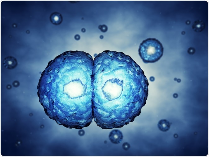
Stem cell discovery began in the 1950s from an unusual source - teratocarcinomas and tumors containing a variety (or heterogeneous mix) of tissue types. These include fully specialized, differentiated structures such as teeth and hair.

nobeastsofierce | Shutterstock
The observation of malignant growth and transplantability suggested the presence of a proliferative (highly dividing) and undifferentiated (non-specialised and capable of producing a range of cell types) cell. This cell was named the embryonal carcinoma (EC) cell.
A pioneering experiment demonstrated that the injection of EC cells in the adult mouse brain resulted in teratocarcinomas; this provided concrete evidence that EC cells can produce all cellular constituents of the teratocarcinoma. Corroborating evidence for their ability to propagate tumors and self-renew was indicated by their ability to be transplanted.
Germ cells and embryonal carcinoma cells
The origins of EC cells are primarily testicular in humans and mice. Leroy Stevens discovered that the teratocarcinomas in 129 strains of inbred mice arose from germ cells, those of the sperm and egg. Paradoxically, germ cells do not give rise to tumors, nor do they differentiate into other cell types.
EC cells exhibited similar morphology (shape) to cells present in the early embryo, which corroborated their germ cell origin. These embryonic founder cells possess the ability to produce the cells of all body lineages, called somatic cells. This property is described as pluripotency, and such cells could be maintained when a sample of the teratocarcinoma was serially transplanted.
Discovering potency
EC cells are like cells in the early embryo that occur prior to gastrulation. EC cells can be expanded into cell line, which was actually achieved in 1970 with cultures from testicular and embryonic teratocarcinomas.
The successful explanation required the use of fibroblasts, which had been inactivated to prevent mitotic division. These cells secrete factors that support the maintenance, proliferation and viability of EC cells.
Furthermore, the EC lines showed variability in their ability to differentiate, an observation which prompted the hypothesis that not all cells were pluripotent; more specifically, there was a difference in the potential of the cell to give different specialized (differentiated) cell types.
This quality is termed potency, of which there are four types, in the hierarchal order:
- Totipotency – the potential to give rise to all the cell types in the embryo and adult (e.g. the fertilized egg), thus a totipotent cell can give rise to the whole organism
- Pluripotency – the potential to give rise to cells of all body lineages, but not a whole organism
- Multipotency – the potential of a cell to give rise to a limited number of cell types in the body
- Unipotency – the ability of a cell to give rise to only a single cell type
From these self-renewing, undifferentiated cells terminally differentiated cells can arise. The scientists Martin Evans and Gail Martin instead focussed on ways to retain pluripotency, which we presently know to be the most potent type of cell. They found that subclones of pluripotent EC cells when grown on a feeder layer, and in the absence of this layer, produced a mixed population of fibroblastic cells (differentiated cells) interspersed with nests of undifferentiated cells.
This finding suggested that EC cells could produce their own feeder fibroblast cells through differentiation and inferred that these must be necessary to maintain the pluripotent nature of these cells. Concurrently, Martin and Evans noticed that the serum in which EC cells were suspended differed in their ability to support EC cell expansion and ability to differentiate. This led to the pluripotency, and subsequently to a reliable culture methodology to produce and propagate EC cells with retained pluripotency.
Stem cells as initators of embryonal growth and development
Further probing of EC cells by Martin and Evans revealed that when EC cells were aggregated, the colonies that arose formed structures called embryoid bodies with specific embryonic identity due to their differentiation features.
The similitude between the two prompted further exploration; to test if the EC cells could contribute to an embryo, the EC cells were injected into blastocysts (pre-embryo structures formed 5-6 days after fertilization) and the outcomes were analyzed.
In most cases, EC cells failed to incorporate into the embryo, some produced tumors or developmental abnormalities. However, some pluripotent EC cell lines colonized the host embryo, and subsequently, chimeras were born.
Chimeras are organisms that contain two different sets of DNA; in this instance, the mice were produced from the DNA encoded in the sex cells and the DNA of the EC cells. This demonstrated that a subset of EC cells could fulfil the function of normal embryo cells and work in the context of the host embryonic environment.
Towards a healthy chimera
However, the proportion of chimeric mice was poor. This finding, coupled with their vast differences in potency between lines, the development of chimeric tumors and changes in their genetic stability, prompted the exploration of another source of pluripotent cells.
A probable explanation for these occurrences was the invariable probability that EC cells carry chromosomal anomalies. All the results suggested that their similarity to embryos were questionable; more specifically, their ability to act in an embryonic-like, pluripotent manner was conditional upon their occurrence in tumors and their inherent genetic abnormalities.
Instead, the embryo itself provided a less problematic source of pluripotent stem cells. Evans and Martins used the same conditions for the optimized harvesting of EC cells on feeder layers in order to successfully harvest undifferentiated cell lines from mouse blastocysts.
The resultant cells bore a functional resemblance to EC cells and could produce teratocarcinomas when they were transplanted into adult mice. This suggested they may have transformed into EC cells.
To eliminate this possibility, in 1984 Evans established that these cells could contribute to healthy chimeras, in which the DNA had been successfully transmitted through the germline (the egg and the sperm). The pluripotent cells, obtainable from an embryo, were expanded through multiple divisions without transforming, retaining their own genetic composition. These cells are presently known as embryonic stem cells (ESCs).
A decade later, primordial germ cells (PGCs) were found to give rise to proliferative stem cells by Matsui. These were termed embryonal germ (EG) cells, and are virtually indistinguishable from their ES cells, apart from their origin. This explained the germ cell origin or teratocarcinomas – the germ cells were induced to convert into pluripotent stem cells in vivo.
Future perspectives: stem cell therapy
The ability for the genetic component of ES cells to be transmitted and retained through the germline offers the opportunity to introduce genetic modifications in mice. They are also amenable to a range of genetic manipulation approaches, and able to be expanded; for example, features that enable separation of cells that have undergone rare event, such as homologous recombination, in which cells undergo some genetic change form exchange between two similar DNA molecules.
In 1989, ES cells with an engineered genetic knockout were successfully generated. This gene targeting of ES cells has since been expanded on. And whilst research in the 1980s and early 1990s focussed on genetic targeting, much of the understanding of why and pluripotency was maintained in ES cells made headway in the late 1990s.
Currently, the molecular mechanisms that underpin the ES cell state have been uncovered. The first reported isolation of human embryonic stem cell lines (hESCs) has shifted the focus away from murine models and allowed exploration of their therapeutic implications. At the moment hESC are indispensable, and the results of hESC‐based clinical trials will set a gold standard for future stem cell‐based therapy.
In 2006, a breakthrough was made by identifying conditions that would permit cellular reprogramming of adult somatic cells to allow them to assume a stem cell-like state. This is now known as induced pluripotent stem cells (iPSCs). Their therapeutic use is potentially greater than that of hESCs, as because these cells can be taken from the patient themselves and then reprogrammed, clinicians can avoid the problem of rejection that is caused by histocompatibility.
The latter refers to the process of the donor possessing different antigens that signal to the recipient host immune system that the cell is foreign and, therefore, lead to an immune response to destroy the foreign cell. This is basically seen as organ rejection.
This additionally avoids the need for immunosuppressive treatment throughout the patient's life to prevent this, and eliminates the ethical implications of hESCs, as iPSCs do not require an embryonic source for production. More research on the topic will definitely bring further progress in stem cell usage.
Sources
- https://www.unmc.edu/stemcells/educational-resources/importance.html
- https://www.ukscf.org/
- http://www.explorestemcells.co.uk/historystemcellresearch.html
Last Updated: May 18, 2023