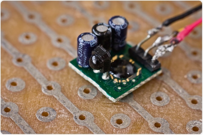
Photodetectors are used in flow cytometers to translate the excited fluorophore light into photocurrent so that it can be digitally stored and analyzed. One of the most common types of photodetectors is photodiodes.

Image Credit: Auhustsinovich/Shutterstock.com
Flow cytometry
Flow cytometry is a technique used for the quantitative and qualitative analysis of cells and particles. It works by analyzing cells as they pass one by one through the flow cytometer’s flow. The cells or particles are tagged with dyed fluorophores. The fluorophores are excited by a light source, such as a laser.
The fluorophores emit photons when excited by the light source, and it is this light that is translated into photocurrent. This allows for both information on the subject that has been tagged with the fluorophore, such as cell size, and for quantification of its components, such as subcellular structures. Since several dyes can be used simultaneously, thus enabling more detailed information on several cellular components.
How do photodiodes work?
Photodiode detectors use cathodes and anodes to transform photons to photocurrent. The photocurrent is an alternative term for the electric current that can be generated from the fluorophore’s photons. Before it arrives at the photodetector, the fluorescent light is divided based on wavelength. This is done by both a combination of dichromatic mirrors and both long pass and short pass filters.
Each wavelength lights up the active area of a chosen photodetector, such as the photodiode. The first step of a photodiode’s function is when the photon hits the photodiode and ionizes the atoms of the detector.
This ionization creates a pair of an electron and a hole within the photodiode, in an area called the depletion region. In the depletion region, the electrons move towards the cathode’s positive potential. The holes move in the opposite direction, towards the anode’s negative potential. This movement creates a photocurrent, which can be moved over to the electronics system.
In most cases, the photocurrent produced by the photodiode is multiplied to an appropriate level. The photocurrent then moves on to the amplifier, where the signal is amplified and transformed to a voltage pulse. Occasionally, a resistor takes this role, instead. The analog to digital converter is responsible for transforming the pulse to a digital number, which becomes the data to be analyzed.
Benefits and limitations of photodiodes
Photodiodes are one of the more established photodetectors that can be used in flow cytometry and are often made of silicon. Other types include photomultipliers and avalanche photodiodes, which are a slightly more advanced version of photodiodes.
These different types of photodetectors have different spectral responses due to different wavelengths. Different types of photodetectors can be used in combination with the same flow cytometer, depending on the light’s wavelength, inity, and pulse duration.
The spectral responses are why photodiodes are typically used for light scatter measurements since photodiodes match better with that light wavelength. This also ties in with the subject matter– standard photodiodes can be used on light scatter at low angles of around 0° to 20°. Because forward light scatter is sensitive to cell size, this can be used on whole cells whereas smaller particles, such as viruses, require larger angles.
Photodiodes are typically used when the forward scatter signal is high, as opposed to when side scatter and fluorescent signals are used (these tend to be weaker than forward scatter signals). However, if the laser or light source directly enters the photodiode, the signal can become overwhelming and result in too much noise. Photodiodes can be used for side scatter and when the fluorescent signals are weaker, but this would require more expensive front end electronics to lower the signal to noise ratio.
While photodiodes are relatively cheap and therefore broadly accessible, they have lower sensitivity than other photodetectors. This is mainly attributed to the fact that photodiodes do not amplify the photocurrent to the same degree other photodetectors, such as photomultipliers.
Flow cytometers with photodiodes are best used in the 350 to 1000 nm range. The maximum current of around 0.5 A/W is at around 900 nm. However, the highest output current is dependent on the power of the laser used to light the sample, the cell’s properties, and the size of the objective lens. While the normal values tend to be in the µA range, which insinuates the top radiant power is in the µW range.
Sources
- Piatek, S., and Hergert, E., 2018. Photodetectors In Flow Cytometers. [online] Photonics.com. Available at: <https://www.photonics.com/Articles/Photodetectors_in_Flow_Cytometers/a64058>
- Thermofisher.com. 2020. Optics Of A Flow Cytometer. [online] Available at: <https://www.thermofisher.com/au/en/home/life-science/cell-analysis/cell-analysis-learning-center/molecular-probes-school-of-fluorescence/flow-cytometry-basics/flow-cytometry-fundamentals/optics-flow-cytometer.html>
- Phinney, D., and Cucci, T., 1989. Flow cytometry and phytoplankton. Cytometry, 10(5), pp.511-521.
- Bakke, A., 2001. The Principles of Flow Cytometry. Laboratory Medicine, 32(4), pp.207-211.
- Thermofisher Scientific. 2020. Electronics Of A Flow Cytometer. [online] Available at: <https://www.thermofisher.com/au/en/home/life-science/cell-analysis/cell-analysis-learning-center/molecular-probes-school-of-fluorescence/flow-cytometry-basics/flow-cytometry-fundamentals/electronics-flow-cytometer.html>
Last Updated: Sep 8, 2020