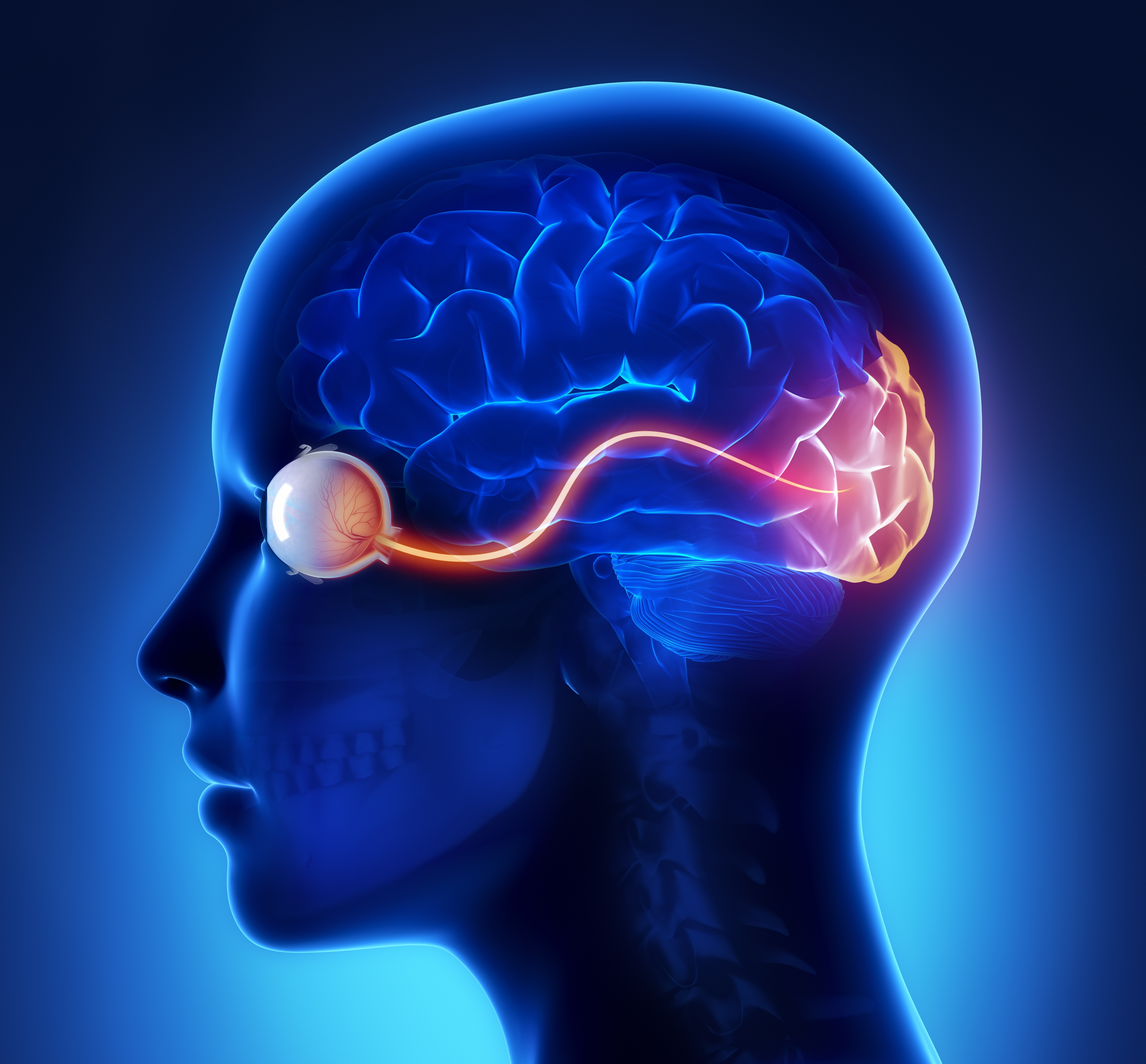
The visual cortex evolved to receive, process, and integrate visual information that enters the brain through the eyes. The information that is processed in the visual cortex is then transferred to other areas of the cortex for further analysis and utilization. Therefore, the neurons in the visual cortex are vital for performing the initial processing of visual information. Activity here feeds into other cognitive processes, meaning that damage to this area can have a profound impact on the human experience.
 Image Credit: CLIPAREA l Custom media/Shutterstock.com
Here, we give a detailed overview of how the neurons in the visual cortex perceive our visual environment.
The structure of the visual cortex
A vast body of research conducted over many decades has resulted in a rich knowledge of the human cortex, the outermost layer of the brain responsible for higher-level processing. The cortex is divided into lobes: the frontal lobe, temporal lobe, parietal lobe, and occipital lobe - each associated with the specialization of different cognitive processes. The occipital lobe, situated at the back of the brain, is the home of the visual cortex and is responsible for visual processing.
The visual cortex itself is further subdivided into five distinct areas based on functional and structural classifications. These five areas are known as V 1-5. Visual information passes from the eyes through to the lateral geniculate nucleus located in the thalamus where it then travels onto the visual cortex.
Scientists theorize that this visual information is first processed in one area, before it is passed on to each subsequent segment that processes the information with an increasing level of specialization. The neurons of the primary visual cortex respond selectively to numerous visual features and factors, such as contours and orientation, motion in a particular direction, and changes in stereoscopic depth.
The specialized neurons of the human visual pathway
Neurons situated within the visual cortex are specialized for their function. In particular, they are differentiated as simple and complex neurons. Mostly located in V1 of the visual cortex, simple cells respond to a specific set of visual features including edges and orientations within a single receptive field.
Complex neurons also respond to edges and orientations, however, studies have shown that they do not represent a single receptive field. Rather, it is the summation of multiple receptive fields that feeds into these simple cells, resulting in a response relevant to an accumulation of visual information. Additionally, complex cells are adapted to respond preferentially to movement in the visual field that is traveling in specific directions.
Other specialized neurons within the primary cortex include end-stopped cells. These neurons are adapted to detecting lines of a certain length and the corners of larger visual shapes. When the line or corner of an image extends beyond a certain point, these cells reduce or cease their response.
Magnocellular, or ‘M’ cells are another type of specialized visual neuron. M cells are large neurons that are found in the retina of the human eye. Studies have suggested that these neurons detect features related to movement, such as direction, location, and speed of a moving object, leading to the conclusion that they are vital to the processing of moving objects.
Smaller neurons, named parvocellular, or ‘P’ cells, are hypothesized to be fundamental for spatial resolution. Studies have shown that these cells are important to processing features such as the color, shape, and size of an object. P cells have also demonstrated their role in color vision.
Kiniocellular, or ‘K’ cells, are small neurons whose role is currently not fully understood. Some studies have suggested that these cells may also be involved in color vision.
Within the primary visual cortex, neurons are arranged into columns with other neurons whose functional properties are similar. Neurons collected into one column might respond to vertical orientation information as a priority, they may also only process information from the contralateral eye.
Neurons in another column, however, may also primarily respond to vertical orientations but from the ipsilateral eye. The information processed in groups of these columns is accumulated into modules, each module contains the information essential for processing one tiny area of the visual field. Therefore, to process an entire visual scene, in any given moment, the visual cortex must combine all this information being generated by each module simultaneously.
While there is a wide body of research on the functioning of the visual cortex and its neurons, there is still much to uncover about how the visual cortex integrates all this information. It is unclear how it brings information together from all areas in order to produce a coherent interpretation of the visual field. Work continues in this area of study and it is likely that over the coming years our knowledge of the intricate workings of the visual cortex will become clearer.
References:
- DiCarlo, J., Zoccolan, D. and Rust, N., 2012. How Does the Brain Solve Visual Object Recognition?. Neuron, 73(3), pp.415-434. https://www.ncbi.nlm.nih.gov/pmc/articles/PMC3306444/
- Goodale, M. and Milner, A., 1992. Separate visual pathways for perception and action. Trends in Neurosciences, 15(1), pp.20-25. https://pubmed.ncbi.nlm.nih.gov/1374953/
- Tran, A., MacLean, M., Hadid, V., Lazzouni, L., Nguyen, D., Tremblay, J., Dehaes, M. and Lepore, F., 2019. Neuronal mechanisms of motion detection underlying blindsight assessed by functional magnetic resonance imaging (fMRI). Neuropsychologia, 128, pp.187-197. https://pubmed.ncbi.nlm.nih.gov/30825453/
Further Reading
Last Updated: Oct 4, 2021