Flow cytometry is a high-throughput method of cell analysis, where cells are passed by an array of lasers and detectors in a single file to quickly determine a range of cellular characteristics and allow rapid cell sorting.
The way in which the laser light is scattered by the cell can be used to infer the current state of apoptosis of the cell, while the combined use of fluorophores allows for the identification of specific structural features inside and on the exterior of the cell by fluorescence microscopy. Other characterization methods such as mass spectrometry may be combined with flow cytometry to allow more refined characterization to be undertaken in situ.

Image Credit: peterschreiber.media/Shutterstock.com
What are circulating tumor cells?
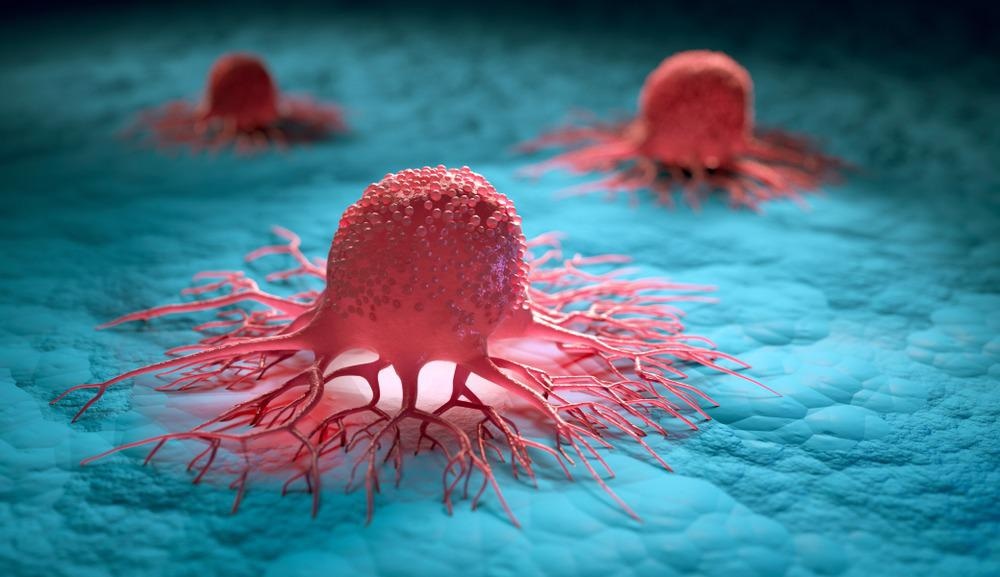
Circulating tumor cells (CTCs) are those that have detached from a tumor and are carried around the body in the blood, potentially engaging in metastasis and developing into a secondary tumor. CTCs are useful prognostic biomarkers that can be used to monitor disease progression and effectiveness of therapy, with the additional advantage of being collected easily and non-invasively for testing via blood sample.
However, even in patients with severe metastatic disease, CTC counts are as low as 1-10 cells per mL of whole blood, compared to around 1 million white and 1,000 million red blood cells. Affinity-based assays towards CTCs are used in CTC testing, with the only FDA-approved methodology for whole blood CTC analysis, CellSearch, involving the separation of CTCs by centrifugation and bonding with tagged magnetic iron nanoparticles to firstly purify the sample. CTC-specific monoclonal antibodies with fluorescent probes are then used to identify and characterize the cells.
Tagging circulating tumor cells
Whole blood analysis by flow cytometry is commonplace, and the technique has also been adapted to CTC analysis by several research groups. The high-throughput nature of flow cytometry aids in the detection of such extremely rare cells, which are usually pre-stained with CTC-specific fluorescent probes to allow easy identification.
Unfortunately, CTCs only have a short half-life of a few hours ex vivo, severely limiting the time available for staining. Further, while CTCs originating from a known source may initially bear known markers for which specific tags may be selected, such markers may be lost during circulation, making tag selection difficult.
Multiple fluorescent tags may be utilized simultaneously during flow cytometry to narrow the range of search parameters filtered. For example, in a paper by Lopresti et al. (2019) the authors utilize multiple fluorescent probes to identify epithelial CTCs by flow cytometry: a CD45 tag as a negative control for rare hematopoietic cells, which bear this antigen; a 4',6-diamidino-2-phenylindole (DAPI) tag to select for nucleated cells, thus removing most white blood cells; and three tags specific to epithelial cells. Additionally, cell size was determined by forward-scattering of light, and as epithelial-derived CTCs are expected to be larger than monocytes and granulocytes this applied an additional filter.
Using this method the group observed that donor blood from individuals without cancer bore far fewer cells of epithelial origin than in those with breast or colon cancer, which had similar rates. The count of CTCs in the blood was also found to be a useful prognostic indicator amongst these and other patients using this method, where those with ten or more CTCs per milliliter of blood had median survival rates of only 2.7 months, compared with 12.5 months for those with fewer than ten.
Epithelial cell adhesion molecule (EpCAM) is the marker most often utilized for the detection of CTCs, though as mentioned these antigens often shed early and become undetectable, and thus other known cancer markers may be utilized. For example, in a paper by Takahashi et al. (2020) markers for HER2 and abnormal chromosome 17 are utilized, both indicators of breast cancer.
Other analytical methods such as mass spectrometry are sometimes incorporated into flow cytometry, and some early studies have utilized mass spectrometry in the single-cell identification of CTCs, though as yet no major studies have incorporated these technologies given the limitations associated with the low population of CTCs in whole blood. Future advancements in CTC enrichment and flow cytometry technology may allow better use of CTCs as diagnostic and prognostic markers in the clinic, replacing the currently employed low-resolution assay.
Continue Reading about Flow Cytometry in Cancer Research Here!
References:
Further Reading
Last Updated: Jan 24, 2022