Adenosine triphosphate (ATP) is a key molecule which upon hydrolysis provides energy to facilitate a variety of cellular processes that are essential for life.
The cell utilizes the energy of ATP hydrolysis in order to drive many non-spontaneous cellular processes. The energy released by breaking the bonds between the phosphate groups can be harnessed to fuel many life sustaining reactions such as:
- Synthesis of biomolecules such as proteins, lipids, DNA and RNA.
- Active transport by pumping ions against a concentration gradient.
- Mechanical work such as muscle contraction, rearrangement of the cytoskeleton and beating of cilia.

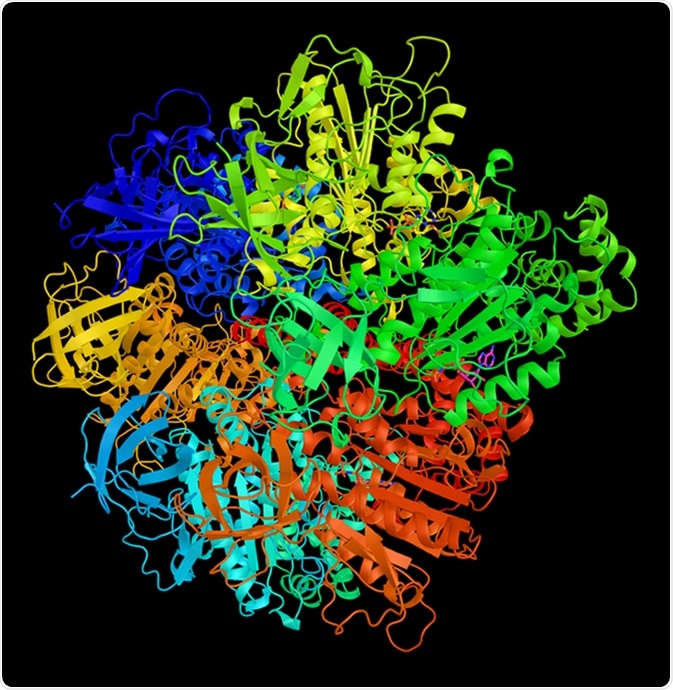
ATPase F1 complex gamma subunit, which forms the central shaft that connects the F0 rotary motor to the F1 catalytic core. 3d rendering. Image Credit: ibreakstock / Shutterstock
ATPases
Although ATP hydrolysis is a favorable reaction, ATP does not breakdown on its own. This is because the activation energy required for the hydrolysis of ATP is high enough that ATP hydrolysis does not take place without an enzyme called ATPase. The lone pair of electrons on the oxygen of a water molecule perform a nucleophilic attack on the terminal phosphate group. However, these electrons have a negative charge and are strongly repelled by the negative charges on the phosphate molecule.
ATP synthase: Structure and Function
ATPases help overcome this repulsion by surrounding the ATP molecule with positive ions that interact with the negative charged ions on the phosphate molecule, allowing hydrolysis to take place. Hence, ATPases lowers the activation energy required for the reaction to occur. Most ATPases leverage the energy released from hydrolysis to either phosphorylate a molecule or change the conformation of ATPase and transport solutes against the concentration gradient.
The different types of ATPases are as follows:
F-ATPases
Reversible ATPases that can use a proton gradient to synthesize ATP or create a proton gradient upon ATP hydrolysis. They are found in bacteria, plants (chloroplasts) and eukaryotes (mitochondria).
V-ATPases
Located in Golgi apparatus, endosomes, lysosomes and vacuoles. They hydrolyze ATP and use the energy for protein trafficking, active transport of metabolites and neurotransmitter release.
A-ATPases
Also reversible ATPases and found only in Archaea.
P-ATPases
Found in bacteria and eukaryotes and responsible for transport of a variety of ions against the concentration gradient using the energy generated by ATP hydrolysis.
ATPase Structure
Most of the ATP is produced in the mitochondria, which comprises of an outer and inner membrane. The space between these two membranes is known as the intermediate space, while the space surrounded by the inner membrane is known as the matrix. The inner membrane also possesses numerous infoldings known as cristae, which contains the enzyme for ATP synthesis.
ATP is generated within the matrix by an enzyme called ATP synthase. It consists of 2 parts:
The F1 portion is located in the matrix that serves as the catalytic site for ATP synthesis or hydrolysis. Whereas the F0 part resides in the membrane and serves as a rotor-like motor and channel for proton transport from intermediate space to the matrix. The F0 subunit consists of 10 c-subunits that form the rotor-like motor, one a-subunit that acts like a proton channel and one b-subunit that extends from the inner membrane into the matrix to stabilize the catalytic subunit of F1 through its contact with the d subunit.
The F1 portion of the enzyme serves as the axle and the catalytic part of the ATP synthase. The axle is comprised of one g and one e subunit which are connected to the c- subunits and extend up into the catalytic subunit that consists of the 3 a and 3 b subunits.
Mechanism of ATPase
The a3b3 hexamer of F1 ATPase are arranged alternately around the g-subunit with the b-subunits being catalytically active. When g-subunit rotates the a3b3 hexamer is held in place by the d and b subunits. Rotation of the g-subunit relative to the fixed a3b3 hexamer causes each b-subunit to cycle through three different conformational states namely bE (empty), bTP (ATP bound) and bDP (ADP bound).
Two alternative schemes have been proposed for the hydrolytic cycle in F1 ATPase. In the first scheme, ATP binds to bE-subunit resulting in a conformational change of the subunit. This produces a clockwise rotation of the g-subunit by 120o. This causes the bDP-subunit into an “open” conformation leading to the release of ADP and Pi. Conformational changes are also transmitted to the bTP-subunit promoting the hydrolysis of bound ATP into ADP and Pi.
In the next step, ATP binds to the bDP-subunit (now empty) and via rotation of the g-subunit results in the release of ADP and Pi from the bTP-subunit and ATP hydrolysis in the bE-subunit. In the final step, ATP binds the (now empty) bTP-subunit, ADP and Pi is released from the bE-subunit and hydrolysis of ATP in the bDP-subunit. In this scheme, the conformational changes induced are a result of ATP binding and not ATP hydrolysis. Also, the bound ATP favors a “closed conformation as opposed to bound ADP and Pi that favor an “open” conformation.
In the second scheme, ATP hydrolysis is directly responsible for the conformational changes induced in the b-subunit. ATP binding to the bE-subunit causes a small conformational change in bDP-subunit resulting in the hydrolysis of ATP at this site. The hydrolysis leads to a large conformational change in the bDP-subunit releasing the products ADP and Pi. The g-subunit rotates 120o clockwise as a result of the conformation change in bDP-subunit causing the ATP-bE subunit to adopt a “closed” conformation. In the next step, ATP binds to the now empty bDP-subunit promoting the hydrolysis and release of ADP and Pi from the bTP subunit. Finally, ATP binds the (now empty) bTP subunit causing the release of ADP and Pi from the bE-subunit bringing the system back to its original state.
The major difference between the two schemes is that ATP binding to the bE-subunit promotes the hydrolysis of the bTP-subunit in the first scheme and bDP-subunit in the second scheme. Also, in the first scheme, two catalytic sites are occupied by ADP and one by ATP. In contrast, two catalytic sites are occupied by ATP and one by ADP in the second scheme.
Sources
- Leslie AGW and Walker JE “Structural model of F1-ATPase and the implications of rotary catalysis. Philos Trans R Soc Lond B Biol Sci. 2000 Apr 2009; 355(1396): 465-471.
- Okuno D, Iino R and Noji H “Rotation and structure of F0F1-ATP synthase” J Biochem. 2011 Jun; 149(6): 655-64.
Last Updated: Feb 26, 2019