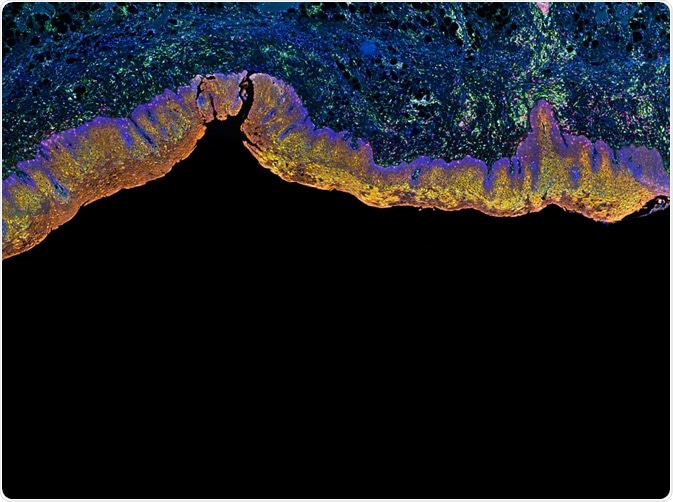
Multi-color immunohistochemistry (IHC) is a technique used to stain multiple antigens within a tissue section. Typical kits are designed for three or more different targets and are used to determine the spatial organization of particular molecules within healthy and diseased tissues.
 Carl Dupont | Shutterstock
Principles of multi-color IHC
Immunohistochemistry is commonly used to visualize the distribution of a target component and its intensity. Routine IHC is limited by the number of colors that can be used, meaning multiple sections must be taken, stained and overlayed. This is not always possible, for example, if there is only a small sample available or a lack of time.
Multi-color IHC solves this problem and allows visualization of tissue architecture, spatial distribution of cellular components, and co-expressed signals. Immunofluorescence has been a favored method to detect several markers in one tissue, but unlike multi-color IHC, it is not permanent. This allows researchers to repeatedly revisit samples without changing the staining results. IHC can also show markers relative to the comprehensive tissue morphology.
Types of multi-color IHC
There are a number of types of multi-color IHC, including brightfield and fluorescence microscopy. Brightfield microscopy represents an elementary form of microscopy which requires chromogens or enzyme pairs to be deposited for multicolor IHC. This is sufficient for distinguishing cell types, but is less apt for intracellular characterization. Counterstaining dyes can be used to enhance multicolor in brightfield microscopy, such as methyl green.
One of the main issues with this type of multicolor IHC is to ensure the enzymes do not cross react. As a result of this, protocols are usually labor intensive and have the risk of colors overlapping. The practicality of exceeding three different colors is limited.
Fluorescence microscopy is a bit more complex, and in the IHC context records the different spectral peaks of light against a dark background, as the fluorophores in the samples are excited by one wavelength.
IHC relies on the binding of an antibody to an antigen of interest, and can be carried out through a direct or indirect process. In direct fluorescence, a fluorophore is conjugated to a primary antibody directly, whereas in indirect fluorescence, the fluorophore is conjugated to an antibody which is specific for the primary antibody.
Several fluorophores are available for IHC, in addition to recently developed quantum dot nanocrystals which have narrower emission peaks, meaning there is less overlap. However, excitation/emission filter sets can physically restrict the ability for multicolor IHC. Typically, it is the filter sets which limit fluorophore markers to three colors, but this can be amended to include more.
How is multi-color IHC applied in research?
Fluorescence multicolor IHC with four different fluorophores have been applied to look at that the tumor environment in gastric cancer. This used the aforementioned indirect labeling method, in turn, applifying the target signal.
In addition, the ability to visualize several immune checkpoints simultaneously in a cancerous tissue can elucidate their expression profiles on immune cells and other cells within a tumor.
Finally, visualizing the co-expression of signaling molecules can be important for furthering our understanding of the cell cycle. The transitional nature of the cell cycle has recently received attention, and the opportunity to simultaneously capture these markers of the cell cycle can help elucidate controls of cycle progression.
Future perspectives
Despite it's successes, multicolor IHC comes with its own limitations. With increased colors in one tissue section, the samples can be difficult to interpret given blending of colors, as often happens because fluorescence is often used. Some types of multicolor ICH also bring the risk of autofluorescence by the tissue itself, which makes visual assessment somewhat difficult.
It has been suggested that confocal laser scanning microscopy can be used, mainly to quantify fluorescence present. Spectral microscopy of this kind can detect the spectra of several markers independently, which prevents them from mixing. This specific procedure has been applied to tonsil tissue, with five different colors.
Last Updated: Jan 3, 2019