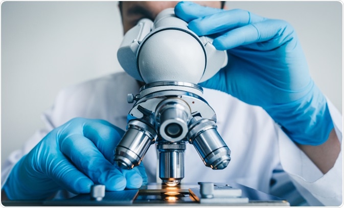
Brightfield microscopy has been widely used for hundreds of years, while reflection contrast microscopy is relatively new technology. As one of the earliest and simplest microscopy techniques, brightfield microscopy is easily accessible but lacks the depth achieved with reflection contrast microscopy.
This article summarizes the similarities and differences of brightfield microscopy and reflection contrast microscopy for different applications.
 Konstantin Kolosov | Shutterstock
Reflection contrast microscopy
Reflection contrast microscopy has a penetration depth of up to 30 µm and a narrow depth of focus, which enables the possibility of 3D reconstructions and imaging living cells.
Advantages
Reflection contrast microscopy operates by suppressing stray reflections and only capturing that which comes from the targeted section. It uses incident light as a light source, and together with a polarizer and a lambda quarter wave plate it polarizes and repolarizes the front lens.
In combination with a decently aligned analyzer, this permits the eyepiece to only receive reflections from the targeted section. Thus. reflection contrast microscopy can be applied to polarizers and wave plates to suppress stray reflections.
Limitations
The image created in reflection contrast microscopy represents only the light reflected from the target. This is fundamentally difficult in biological samples because biological objects have low reflected light intensities less than 1%, whereas glass-air interfaces have reflection intensities of 4%.
Brightfield microscopy compared to reflection contrast microscopy
Brightfield microscopy can be used to visualize cells by contrasting the dark specimen with bright light. Brightfield microscopy uses transmitted light as a light source, compared to reflection contrast microscopy which uses incident light. The light passes through the specimen and is collected by the objective lens, which magnifies the light and further transmits it to an oracular lens through which the image is viewed.
To view particular structures, staining, such as fuchsin and methylene blue are used. In reflection contrast microscopy, interference created by stray reflections are removed by using added machinery to a fluorescence or light microscope.
Brightfield microscopy can also be applied to living cells and for 3D reconstruction when coupled with imaging accessories. It typically requires staining of the cells which can be quite general in comparison to the higher specificity immunocytochemistry or in situ hybridization achieved using reflection contrast microscopy. However, staining in brightfield microscopy cannot be carried out on live cells.
Applications of brightfield and reflection contrast microscopy
Brightfield microscopy can be used to generate high resolution images, such as several organisms interacting or larger subcellular structures, such as cell borders and nuclei.
Reflection contrast microscopy, is better suited for subcellular imaging or imaging of the components of the extracellular matrix. However, in cells with intrinsically colored subcellular components, such as chloroplasts in plant cells, brightfield microscopy can be used. The magnification with brightfield microscopy is limited to the wavelength spectra of visible light.
One of the primary benefits of brightfield microscopy is that it is easy to apply. For example, brightfield microscopy is commonly used in criminal investigations to image hair samples and identify food remains.
Brightfield microscopy is very user friendly, and is often the microscope students are initially familiarized with. Reflection contrast microscopy is more complex, requiring adaption equipment and more complex sample preparation before imaging. However, reflection contrast microscopy can be carried out using a fluorescence or confocal laser scanning microscope, provided the adaption equipment is applied.
Although both methods require a control, it is slightly more critical in reflection contrast microscopy because the control can determine if the antibody is bound to the correct substructures.
Further Reading
Last Updated: Apr 1, 2019