The Intestinal Stem Cell Niche
The stem niche of the intestine is located within the crypts of Lieberkühn and provids physical protection to stem cells. These cells are constantly replenishing themselves, and the surface epithelium is replaced every 5 days. The other parts of the intestine is composed of villus and crypts to maximize surface area, while there is no villus in colon.
The stem cells within the intestine have four potential cell fates: absorptive, goblet, enteroendocrine and paneth cells. The most common cell type within the colon are the absorptive cells.

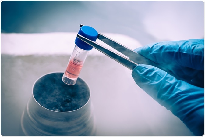
A Liquid Nitrogen bank containing suspension of stem cells. Cell culture for the biomedical diagnostic. Image Credit: Elena Pavlovich / Shutterstock
The Different Types of Stem Cell Within the Intestine
There are also two different types of stem cells within the intestine:Lgr5+ cells, which replicate constantly, have high telomerase activity, produce the crypts; BMI1 cells, which divide under extreme stress, such as removing colon during surgery. BMl1 cells are very resistant to radiation and activating growth factors, such as Wnt.
Factors Controlling the Development of Stem Cells in the Colon
Niche and environmental factors
Niche and environmental factors ensure the precise regulation of differentiation. There are many signaling pathways that maintain and differentiate cells, including the Wnt/β-catenin, transforming growth factor (TGF), bone morphogenic protein (BMP), Hedgehog (Hh), and Notch signaling cascades. Transcription factor Wnt is produced by myofibroblasts at the bottom of the crypts and signals proliferation. Hh is produced at the top of the crypt and inhibits the transcription of Wnt genes. BMP inhibits both Wnt and Hh, and both Noggin and Gremlin are produced by cells within the crypt to prevent the function of BMP. Therefore, the differentiation of the colon is highly controlled by a network of signals which balance the action of Hh and Wnt and therefore control the fate and development of a cell.
Regional segregating factors
The location of individual cells is determined by regional segregating factors, such as Ephrin B1/B2, which control the localization and migration of cells. Different Ephrin types are produced in different parts of the crypt, stimulating the migration of cells depending on the receptors on their surface. The location of the cell determines which cell type it differentiates into. For example, paneth cells do not have Ephrin receptors and do not migrate up the crypt. The signal at the crypt base stimulates the formation of paneth cells. Notch receptors and Jagged ligands also lead to lateral inhibition between adjacent cells. Notch activation drives the differentiation of absorptive cells, while inhibition drives the differentiation of secretory cells. Therefore, the fate of the cell is determined by adjacent cells and signals from other cell types.
Recreation of the Intestinal Niche
Studies have identified a method to maintain crypts within a culture: cells are kept on basement membrane and provided with Noggin, Wnts, and R-spondin growth factors. This system can be used for tissue engineering, regenerative medicine, model development, and toxicology assays. However, lineage development is limited and certain cells which are usually found within the crypt are lacking.
Cell growth and differentiation in the Intestinal crypt
Last Updated: Oct 29, 2018