Skip to:
What are tumorspheres?
Numerous studies have pointed out that cancer stem cells (CSCs) play an important role in cancer progression, metastasis, relapse, and resistance to therapy. Study of these CSCs can shed light why many existing therapeutic regimens eventually fail.

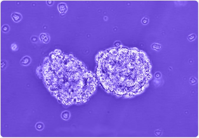
Tumorspheres. Image Credit PLOS https://doi.org/10.1371/journal.pone.0063519
The proliferation of CSCs in non-adherent, serum-free conditions gives rise to fused cells that cannot be identified individually. Such solid and spherical formulations of fused cells are called tumorspheres. The size of tumorspheres usually ranges between >50 µm and 250 µm.

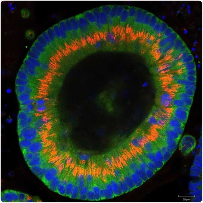
Colon cancer cells grow into three-dimensional organoids in the culture dish. Dr. Joseph Regan / Charité - Universitätsmedizin Berlin
Tumorspheres are used to determine the percentage of cancer stem cells present in a tumor population, but also for studying the ability of such cells to regenerate themselves. In addition, cultivation of tumorspheres can also be beneficial in determining a potential anti-cancer agent for therapeutic use.
Tumorsphere formation assay
Cell cultures are usually used to study tissue morphology, mechanisms involved in the disease, and effect of drugs on the diseased state. However, traditional systems such as 2-dimensional (2D) cell cultures cannot yield large number of CSCs and have other disadvantages such low efficiency, spontaneous fusion among spheres creating large spheres that are unstable, time consuming, and high costs.
On the other hand, 3D cultures are slowly gaining popularity due to the possibility of optimizing culture conditions in vitro and producing CSCs that have spheroidal structures, resembling in turn a solid tumor. The aforementioned features of 3D cultures facilitate the study of different drug therapies and biomarkers.
Scientists have been using different microfluidics methods to study CSCs due to their high throughput and precision. Pang and collaborators developed a microfluidic device that forms spheres from single cells and helps in analyzingdrug-resistant qualities of the tumorsphere. The micro-device contains multi-obstacle architecture-looking (MOAL) microstructural matrices along with micro-well arrays.
In short, these researchers studied glioblastoma cells in addition of the anticancer drug vincristine, and were able to observe different sizes of the glioblastoma cells. It was noted that the tumorspheres acted like stem cells and their resistance to vincristine and other growth patterns revealed the heterogenic capability of CSCs.

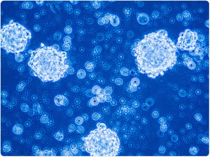
Glioblastoma stem cells organized in tumor niche formation. Image Credit: Anna Durinikova / Shutterstock
Technical notes on tumorsphere cultivation
To prevent cell aggregation during tumorsphere cultivation, it is important to use a culture with low attachment surface – such as 1% agar, agarose gels, 0.5-1% growth-factor reduced Matrigel, or polyhydroxyethylmethacrylate. Adding natural polymers to the culture system prevents sedimentation of the tumorspheres, but does not disturb their inherent properties or ability to proliferate.
In addition, the cell preparation should be filtered through a 40µm cell strainer to obtain a cell density that is less than ten cells per µL. Although tumorspheres cultivation is carried out in a serum-free environment to prevent cell aggregation, there are studies that have reported that a small concentration of serum sometimes aided in tumorsphere formation.
The number of tumorspheres are counted by recording the diameter of the tumorspheres, and expressed as size or total number of formed tumorspheres. This is important, as the number of tumorspheres can be employed to characterize the population of cancer stem and/or progenitor cells both in vitro and in vivo.
What are the types of cancer stem cell (CSC) tumorspheres?
Self-renewing CSCs are required for the growth of any kind of tumor. For instance, human breast cancer cell lines contain these self-renewing CSCs, identified as tumor markers, thereby making them ideal models for developing and testing progenies that are phenotypically diverse, as well as their response to cancer therapy.
Similarly, studies have observed that pancreatic cancer can be identified by c-Met, a tumor marker that is the responsible for the growth of pancreatic tumors. Equipped with such information, researchers can develop tumorspheres in vitro and study their characteristic properties, such as resistance to chemotherapy (as seen in castration-resistant prostate cancer).
The reticent property of this kind of prostate cancer is attributed to the presence of high concentration of stemness genes such as glioma-associated oncogene homolog 1 (Gli1), ATP Binding Cassette Subfamily G Member 2 (ABCG2), BMI1 (a core component of polycomb repressive complex 1), along with overexpressed CD44.
Tumorspheres for screening anti-CSC drugs
With treatment resistance and metastasis linked to CSCs, a number of studies are exploring the possibility of using CSCs for discovering new therapeutic strategies. A recent study in Spain by Pomares and coauthors on non-small cell lung cancer (NSCLC) from eight resected patients and twelve cell lines assessed the CSCs properties of tumorspheres in vitro and in vivo.
Non-obese diabetic (NOD) and severe combined immunodeficient (SCID) mice were given select drugs by the intraperitoneal route after tumor was induced by the tumorspheres obtained from NSCLC patients and cell lines. Results demonstrated lung tumorspheres to be highly resistant to some chemotherapy agents, but also showed the higher cytotoxic potential of some novel drugs.
Tumorsphere cultivation was carried out in a phase I immunotherapy trial by Wang et al that enrolled previously untreated patients with hepatocellular carcinoma (HCC). The patients were treated with autologous dendritic cells derived from patient tumorspheres. In short, this trial demonstrated a 100% efficiency rate with respect to establishing short-term tumor cell lines from resected HCC in comparison to standard tissue culture techniques.
In addition, there was no significant toxicity observed, nor was there worsening of any parameters related to hepatic functions. Such trials hold the promise of producing patient-specific products through this technique in the near future.
What is the future of tumorspheres?
For a successful cancer therapy, it is important to target CSCs. Tumorsphere assays provide a reliable platform to develop anti-CSC drugs, as well as CSC-targeted immunotherapy. Compared to 2D monolayer culture methods, tumorsphere cultivation can provide more definitive biological answers and therapeutic solutions to many challenges that medicine faces during cancer immunotherapy.
Sources
- Lin D et al. (2019). Screening Therapeutic Agents Specific to Breast Cancer Stem Cells Using a Microfluidic Single-Cell Clone-Forming Inhibition Assay. Advanced Science News. Small journal. https://doi.org/10.1002/smll.201901001
- Johnson S et al. (2016) In vitro Tumorsphere Formation Assays. Bio-Protocol. https://www.ncbi.nlm.nih.gov/pmc/articles/PMC4972326/#
- Zhu ZW et al. (2017). A novel three‑dimensional tumorsphere culture system for the efficient and low‑cost enrichment of cancer stem cells with natural polymers. Experimental and Therapeutic Medicine. https://doi.org/10.3892/etm.2017.5419
- Kapałczyńska M et al. (2018). 2D and 3D cell cultures – a comparison of different types of cancer cell cultures. Archives of Medical Science. doi: 10.5114/aoms.2016.63743
- Pang L et al. (2019). Single-Cell-Derived Tumor-Sphere Formation and Drug Resistance Assay Using an Integrated Microfluidics. Analytical Chemistry. DOI: 10.1021/acs.analchem.9b01084. pubs.acs.org.secure.sci-hub.tw/doi/pdf/10.1021/acs.analchem.9b01084
- Lee CH et al. (2015). Tumorsphere as an effective in vitro platform for screening anti-cancer stem cell drugs. Oncotarget. doi:10.18632/oncotarget.6261. https://www.ncbi.nlm.nih.gov/pmc/articles/PMC4811455/
- Fillmore CM et al. (2008). Human breast cancer cell lines contain stem-like cells that self-renew, give rise to phenotypically diverse progeny and survive chemotherapy. https://doi.org/10.1186/bcr1982
- Li C et al.(2011). c-Met Is a Marker of Pancreatic Cancer Stem Cells and Therapeutic Target. Gastroenterology. https://doi.org/10.1053/j.gastro.2011.08.009
- Zhang L et al. Tumorspheres derived from prostate cancer cells possess chemoresistant and cancer stem cell properties. Journal of Clinical Research and Clinical Oncology. https://link.springer.com/article/10.1007/s00432-011-1146-2
- Pomares AH et al. (2019). Lung tumorspheres as a drug screening platform against cancer stem cells. Annals of Oncology. https://oncologypro.esmo.org/
- Lee CH et al. (2016). Tumorsphere as an effective in vitro platform for screening anti-cancer stem cell drugs. Oncotarget. doi: 10.18632/oncotarget.6261
- Wang X et al. (2015). Phase I trial of active specific immunotherapy with autologous dendritic cells pulsed with autologous irradiated tumor stem cells in hepatitis B-positive patients with hepatocellular carcinoma. Journal of Surgical Oncology. doi: 10.1002/jso.23897. https://onlinelibrary.wiley.com/doi/full/10.1002/jso.23897
Further Reading
Last Updated: Aug 18, 2023