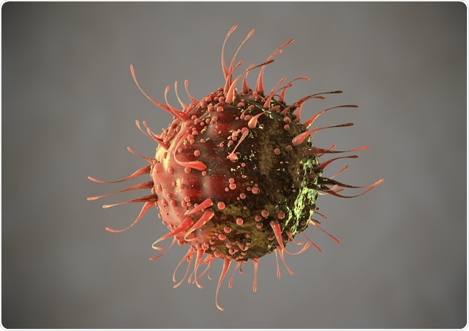
In this case study, researchers at the Weizmann Institute of Science in Israel developed and optimized an imaging flow cytometry (IFC) protocol to provide a high-throughput, quantitative and highly robust method for studying viral infection and virus-host interactions.
The researchers were able to simultaneously monitor diverse cellular processes including the generation of viral factories, transport, the fusion of replication centers, accumulation of viral progeny and changes in morphology.
Skip to:

BertrandBlay | Shutterstock
Obtaining a Full Picture of Viral Infection is Challenging
The heterogeneity of viruses and their hosts means the best way to achieve a full picture of viral infection is to study large populations of infected cells. However, information on variation in infected cells is lost when performing bulk measurements.
Since the discovery of the giant Mimivirus, the majority of what is understood about its infection cycle has been achieved using sophisticated microscopy methods. Although these provide highly detailed structural information, they are limited by their small field of view, which only allows a small number of cells to be imaged at once.
Minsky and team wanted to complement the existing methodologies by developing a high‐throughput method that would quantify the progression of infection and provide accurate time windows for various steps in the replication cycle.
Using Imaging Flow Cytometry to Study Mimivirus Infection in Amoeba
In order to explore the temporal progression of events in infected cells, Minsky and team used imaging flow cytometry (IFC) to monitor the changes in Acanthamoeba polyphaga (amoeba) cells that had been infected with the giant Mimivirus.
Although the team chose to focus on this virus, they say that the IFC platform can be used to study the infection cycles of other viruses.
To study viral infection using IFC, the researchers first needed to develop methods that would preserve the morphological features of adherent amoeba cells before they were detached and analyzed in suspension. They also needed to identify IFC parameters that would best inform on important infection cycle events.
Once the protocol was optimized, the team were able to obtain time windows for the various cellular processes and use IFC to assess the effects of disturbances such as oxidative stress and cytoskeletal disruptors on infection.
By aligning the processes along a timeline, the researchers were able to assemble them into three main temporal phases, which were 0–4 h PI, 4–6 h PI, and 6 h PI to cell lysis.
The study found that mild oxidative stress slowed various stages of virus production, although infection processes did eventually happen at similar amplitudes.
“In this study, we demonstrate that IFC can be used to examine temporal and amplitude variation across infected cell populations for key events in cell reprogramming and viral production,” writes the team in the journal Cytometry.
The Role the Cytoskeleton Plays in Infection
Using imaging flow cytometry to examine specific aspects of virus-cell interactions, the team next asked whether and how cytoskeletal changes might contribute to infection.
When the researchers monitored microtubules and actin microfilaments, they found that both undergo major structural transformations as the cells round up and become immobile during the second infection phase at 4–6 h PI. They propose that translocation of microtubules to the cell periphery may make room for the viral factory (VF) to mature and expand in the cell center and also allow for massive production of virions.
“Notably, such a microtubule rearrangement may be an outcome of changes in the actin network due to viral infection, and thus microtubules may not be direct or active participants in Mimivirus infection,” say Minsky and colleagues.
To find out whether actin microfilaments are required for infection, the researchers disrupted the filaments using latrunculin‐B. They then observed defects in the fusion of replication centers into a mature viral VF and in the production of new virions.
Although the scientists focused specifically on the role of the host–cell cytoskeleton in Mimivirus infection, they say the method could be applied to many other viral or cellular factors that potentially affect the progression of Mimivirus.
Future Research
Further research will now be needed to investigate how Mimivirus utilizes the Acanthamoeba cytoskeleton and shed light on additional fundamental issues of the viral infection cycle.
Finally, they propose that the imaging flow cytometry platform be used to study the infection cycles of other viruses, especially other recently discovered VF‐forming giant viruses, to analyze and quantify viral–host interactions and to explore the progression of infection under different perturbations.
“Through this report, we demonstrate that IFC offers a quantitative, high‐throughput, and highly robust approach to study viral infection cycles and virus–host interactions,” concludes the team.
Source
Minsky, A., et al. (2019). Kinetics of Mimivirus Infection Stages Quantified Using Image Flow Cytometry. Cytometry. https://doi.org/10.1002/cyto.a.23770
Further Reading
Last Updated: Sep 9, 2019