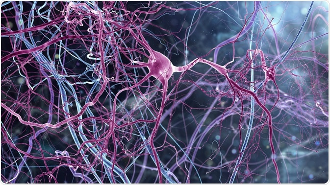
Neuroepithelial cells are stem cells that differentiate into neurons and glia, essential components of the human central nervous system, following the process of neurogenesis.

Image Credit: Whitehoune/Shutterstock.com
Also known as neuroectodermal cells or neural stem cells, neuroepithelial cells are vital to the development of the structure of the brain.
Below we discuss how the cells form the brains of human fetuses, and how they then continue their vital work in supporting the process of neurogenesis in adult brains, which is related to cognitive functions such as learning and memory. It is also associated with the neural repair which has led to connections being made with neuroepithelial cells and neurodegenerative disorders such as Alzheimer’s disease, Huntington’s disease, and Parkinson’s disease, as well as schizophrenia. Therefore, the role of neuroepithelial cells in neurological diseases is also explored.
Neuroepithelial cell proliferation in embryos
Neuroepithelial cells are vital from the very first stages of embryonic development. Two weeks after fertilization the blastocyst morphs into what is known as the embryonic disk. This disk splits into three distinct layers, the ectoderm, mesoderm, and endoderm.
Following differentiation, the mesoderm forms a temporary backbone known as the notochord, this activity releases a chemical that triggers the formation of the neural plate as surrounding undifferentiated ectoderm cells begin to thicken as a result of mitosis of the neuroepithelial cells. Neuroepithelial cells line the lumen of the newly formed neural plate, which then folds first into a neural groove and then into a neural tube.
From inside the tube, the neuroepithelial cells generate more neuroepithelial cells through a process of symmetric proliferative divisions. Following this, the cells give rise to non-stem cell progenitors, creating the two cell types that construct the majority of the human nervous system, the neurons, and the glia.
The rest of the components of the central nervous system (CNS) are derived from the other two layers, with the astrocytes and oligodendrocytes arising from the ectoderm, and the bone marrow and microbial cells arising from the mesoderm.
This process of converting neural stem cells into different CNS cells is essential to developing a healthy brain. Human brains have numerous specialized areas of function, which has led to the need for very specific kinds of neurons.
The hippocampus, which mediates memory and spatial navigation, needs 27 types alone. The utilization of neuroepithelial cells to fuel neurogenesis has evolved to equip the brain with the tools it needs to create all the neural cell types it relies on for its diverse set of complex functions.
Adult neurogenesis
In the adult brain, neuroepithelial cells continue to play a vital role in supporting various CNS functions by generating the necessary neurons and glia. Neuroepithelial cells can give rise to these cells through the process of neurogenesis.
In the adult brain, these cells are found in the senate gyrus of the hippocampus, the olfactory bulb, and the subventricular zone.
In the adult brain, the process of neurogenesis involves the neural stem cells generating radial glial cells, which then differentiate into neurons and glia. The process involves two kinds of mitotic division, asymmetric differentiating division, and symmetric prolific division.
With asymmetric differentiating division, the outcome is the creation of two different cell types, whereas with the symmetric prolific division the two resultant daughter cells are identical. The orientation of the mitotic spindle governs whether the neuroepithelial cell will undergo symmetrical or asymmetrical division.
When the spindle is located in the posterior or anterior area, asymmetric division occurs, whereas when it is in the center, a symmetric division is stimulated. Mitosis can form either more neuroepithelial cells, radial glial cells, or progenitor cells.
When the latter are formed, they are then converted into either neurons or glial cells, using extracellular trophic factors such as ciliary neurotrophic factor (CNTF), cytokines, or neuregulin 1 (NRG1) to decide which of the cell types they will differentiate into.
Many genes, external stimuli, and regulatory pathways of the CNS govern the process of neurogenesis, which helps to fine-tune the workings of the newly created neurons.
Looking at neuroepithelial cells to understand the disease
Throughout our lives, new neurons and glia are needed for physiological brain functions, and abnormalities in neurogenesis are, therefore, associated with many neuropsychiatric diseases.
For example, the olfactory epithelium contains neural stem cells and progenitors, and therefore plays a role in lifelong neurogenesis and regeneration. Recent studies have uncovered that useful disease models can be created through the study of olfactory epithelium tissue, which has given rise to a greater understanding of the alterations in neurodevelopment, gene/protein expression regulatory pathways, and stress response observed in schizophrenia.
Researchers are confident that further study of neuroepithelial cells of the olfactory epithelium would be useful in studying the molecular mechanisms implicated in schizophrenia. What’s more, the study of neuroepithelial cells in various brain tissues could also help to uncover the intricate mechanisms of other neuropsychiatric disorders.
Sources:
Braun, S. and Jessberger, S. (2014). Adult neurogenesis: mechanisms and functional significance. Development, 141(10), pp.1983-1986. https://dev.biologists.org/content/141/10/1983
Lavoie, J., Sawa, A. and Ishizuka, K. (2017). Application of olfactory tissue and its neural progenitors to schizophrenia and psychiatric research. Current Opinion in Psychiatry, 30(3), pp.176-183. https://www.ncbi.nlm.nih.gov/pmc/articles/PMC5471112/
Mitchell, B. and Sharma, R. (2009). The nervous system. Embryology, pp.59-62. https://www.sciencedirect.com/science/article/pii/B9780702032257500130
Wang, M., Zhang, L. and Gage, F. (2019). Modeling neuropsychiatric disorders using human induced pluripotent stem cells. Protein & Cell. https://link.springer.com/article/10.1007/s13238-019-0638-8
Yau, S., Li, A. and So, K. (2015). Involvement of Adult Hippocampal Neurogenesis in Learning and Forgetting. Neural Plasticity, 2015, pp.1-13. https://www.ncbi.nlm.nih.gov/pmc/articles/PMC4561984/
Further Reading
Last Updated: Jan 30, 2020