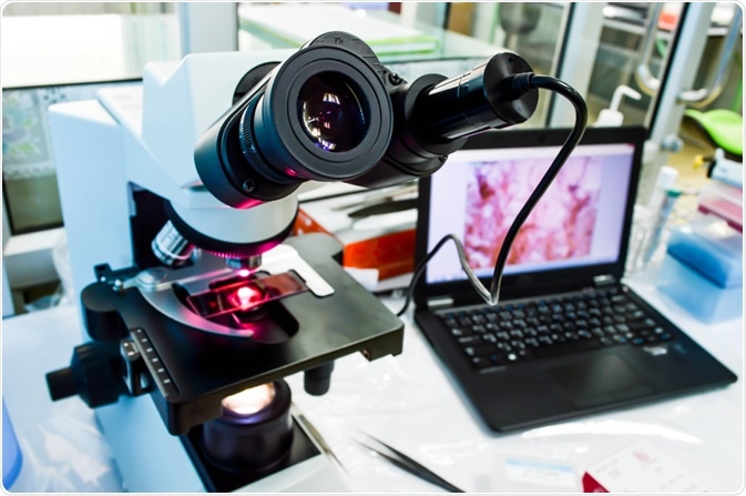
The practice of digital pathology encompasses the sharing and management of digital imagery taken of microscope slides. The process, enabled by computer technology, allows scientists across the world to share and develop their knowledge surrounding the pathology of numerous diseases.

Image Credit: Pakpoom Nunjui/Shutterstock.com
While digital pathology has seen rapidly increasing widespread adoption over the last decade, the idea is not new. The concept of digital pathology dates back over a century, to when scientists first developed specialized equipment to transfer images generated from microscopes onto photographic plates so that images could be retained, referred to at a later date, and shared with other scientists to help gain insights into pathology.
It was roughly 50 years ago that scientists began sharing images between remote locations, representing the emergence of telepathology, practicing pathology at a distance. Limitations in technology have prevented the method from realizing its full potential until the last 10 years.
The last decade has seen a shift from analog to digital in all areas, which has facilitated the emergence of digital pathology. While whole-slide imaging (WSI) scanners entered the mainstream 20 years ago, it has not been until now that they have undergone vast improvements resulting in equipment that can digitize an entire glass slide, making a significant impact on the field of pathology.
Along with advances in equipment, computational technology and storage have also undergone significant improvements, which has also helping to foster the widespread adoption of digital pathology.
The progression of whole-slide imaging, the increased speed of networks, and the cheaper, more accessible storage solutions have resulted in digital pathology establishing itself in modern clinical practice, playing a vital role in the advancement of our knowledge of the nature of the disease.
How does modern digital pathology work?
In modern digital pathology, several basic steps are observed by pathologists and scientists who are involved in the practice. First, information is acquired, which means the digitization of slides as well as related data. To digitize microscope slides technicians use a scanning device to capture the imagery.
Today, labs are mostly relying on automated digital pathology scanners to capture the image information available within the entire glass slide. Images can be recorded in both bright fields and fluorescent conditions while maintaining magnification levels comparable to those achieved by the microscope.
Next, this information is entered into a digital management system where it can be shared, analyzed, and interpreted, all in a digital environment. Scientists around the world using digital pathology software applications can access this data via shared networks.
Automated analysis tools are also included in these software packages that can help speed up the analysis and interpretation of biomarker expressions within samples.
The future of digital pathology
Digital pathology has become a commonplace practice in labs around the world, growing in popularity as advancements in technology have improved its offering and have made it more accessible. It is predicted that this is just the beginning and that digital pathology will continue to evolve and provide enhanced solutions and more benefits.
Scientists are currently working on combining the advances being made in artificial intelligence and machine learning with digital pathology. This would enable image-based diagnosis, which may speed up and improve the accuracy of the diagnosis of several diseases.
Overcoming challenges
While the future looks bright for digital pathology, for it to meet its full potential, several challenges are facing digital pathology which must first be addressed.
The first major issue is that to adopt digital pathology, scientists who are likely to be more familiar in working with the tissue samples themselves, and analyzing them using traditional methods may be reluctant to switch to a new, digital method. For many, this may require retraining, which can be off-putting to staff as well as employers who may be avoidant of allocating time and resources to training.
The field of pathology, in particular, is an area of science that has followed a rigid protocol for many years, leading to scientists becoming experts in a strict set of standard procedures. Switching the entire workflow can, therefore, be intimidating. For digital pathology to truly succeed it must be combined with comprehensive retraining strategies that allow scientists to adapt to the new status quo.
Also, digital pathology faces technological challenges. The use of low-quality computer hardware can lead to inaccurate analysis and even diagnosis. Also, with the high volume of slides produced by the average pathology lab each day, a large capacity is needed to process and store all these images. This means that setting up digital pathology practices can incur large upfront costs, making its adoption somewhat unappealing.
Finally, digital pathology is suffering from a lack of standards and regulations. These need to be set to ensure that the quality and reliability of processes are the same across labs. Standard processes also need to be agreed upon to ensure uniformity of data collected across locations, so that it is easily shared, interpreted and analyzed by different teams.
Sources:
- de Araújo Novaes, M., 2020. Telecare within different specialties. Fundamentals of Telemedicine and Telehealth, pp.185-254. https://www.sciencedirect.com/science/article/pii/B9780128143094000100
- Digital Pathology Market Size, Share & Trends Analysis Report By Product, By Application (Drug Discovery & Development, Academic Research, Diagnosis), By End Use (Hospitals, Clinics), And Segment Forecasts, 2020 - 2027. Available at: www.grandviewresearch.com/.../digital-pathology-systems-market
- Niazi, M., Parwani, A. and Gurcan, M., 2019. Digital pathology and artificial intelligence. The Lancet Oncology, 20(5), pp.e253-e261. www.thelancet.com/.../fulltext
- Pantanowitz, L., Sharma, A., Carter, A., Kurc, T., Sussman, A. and Saltz, J., 2018. Twenty years of digital pathology: An overview of the road traveled, what is on the horizon, and the emergence of vendor-neutral archives. Journal of Pathology Informatics, 9(1), p.40. https://www.ncbi.nlm.nih.gov/pmc/articles/PMC6289005/
Further Reading
Last Updated: Mar 30, 2020