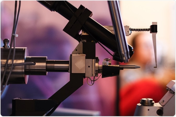
X-Ray crystallography is a tool used to provide structural information about molecules. The technique was developed in 1912 by William Henry Bragg and William Lawrence Bragg (a father and son team who won the 1915 Nobel Prize in Physics for their work in the field), who built upon earlier work by Max von Laue.
Von Laue discovered that by shining X-rays through a copper sulfate crystal onto a photographic plate, diffraction spots that related to the crystalline structure of the sample were produced.
Skip to:
 Gregory A. Pozhvanov | Shutterstock
An introduction to X-ray crystallography
X-ray crystallography uses electromagnetic radiation (specifically, X-rays) to determine the molecular and atomic structure of a crystal. The structure of the crystal causes the X-rays to diffract in specific directions. Through analysis of the intensities and angles of these beams, the position and arrangement of electrons within the crystalline structure can be determined.
A three-dimensional picture of the electron densities can then be produced. Information such as the mean position of atoms within the structure, covalent bonding between them, and their crystallographic disorder can then be determined, which represents the three-dimensional structure of a molecule.
The reason X-rays are used in this process is because the clouds of electrons are at the same scale as the X-ray radiation wavelength. This means that the radiation is deflected and scattered by the electrons of the atoms in the crystal. The deflected X-ray beams produce a scattering distribution which is proportional to the scattering angle. Bragg’s law is used to describe this.
Since many diverse types of structure can form crystals, X-ray crystallography can have many research applications. The substances that can be analyzed by this method include salts, minerals, metals, semiconductors as well as biological compounds including proteins, nucleic acids, and vitamins.
The most difficult part of the process is growing a perfect crystal, as this is needed to provide accurate information about the sample. Some macromolecules, especially those with a high atomic weight such as membrane proteins, can be difficult to crystallize.
Many different fields of study including biology, chemistry, and geology have found uses for this powerful yet simple technique.
Seeing Things in a Different Light: How X-ray crystallography revealed the structure of everything
Methodology
An X-ray crystallography machine works by utilizing a four-circle diffractometer. This works by rotating the crystal and the deflector between the X-ray source and the screen. The screen receives the X-rays which have passed through the crystal.
X-Ray crystallography experiments are broken down into four steps:
- Protein crystallization
- Production of a diffraction pattern
- Analysis of the diffraction pattern to produce an electron density map
- Determination of the protein structure.
As has been mentioned, the most difficult part of the process is achieving a perfect crystal structure for analysis. As the position of electrons must be mapped accurately, it is important that the structure be flawless.
A diffraction pattern is formed on the screen by the X-rays which have been absorbed by the atoms in the crystal, which leave behind dark diffraction spots. Their density varies with the amount of interference between the diffracted electrons at each point. These spots represent accurately the electron density, which can be mapped.
Once the electron density map has been made, the analysis of the crystallographic data is relatively straightforward. It does, however, require complex mathematics to make sense of the information. In the early days of X-ray crystallography, these calculations were done by hand, but now computers are used to perform them.
The calculation used is called the Fourier transformation. This calculation transforms the data into a three-dimensional representation of the atomic or molecular structure of the sample molecule or material.
Applications of X-ray crystallography
X-ray crystallography is used to analyze many different molecules and has been used in many famous projects in the fields of organic and inorganic chemistry. Early structures which were resolved using the technique were simple crystals, including quartz and salt.
One of the most well-known of these was the determination of the double-helical structure of DNA by Franklin, Watson, and Crick in 1953. Other important molecules whose structures were identified include Vitamin B12, insulin, and penicillin.
As well as the analysis of organic molecules (proteins, vitamins, nucleic acids) and inorganic molecules and structures, X-ray crystallography has been used to develop novel materials in both material and life sciences.
Summary
X-ray crystallography is still one of the best methods for the structural analysis of many substances. It remains a powerful, simple, and reliable technique that is used by laboratories across the world.
There are numerous studies that X-ray crystallography can provide unique insights for that other methodologies cannot, but it does have some disadvantages compared to these other techniques, which is why it should always be used as part of a suite of analytical methods.
Further Reading
Last Updated: Oct 14, 2019