The analysis of proteins and nucleic acids within large, intact organs is a very difficult process. Prior to the development of the CLARITY technique in 2013, such analytical methods involved sectioning and restructuring and were thus limited to small and thin tissues.
 Johan Swanepoel | Shutterstock
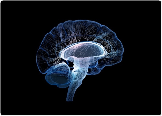
The brain contains 86 billion neurons, which are assembled into very complex circuits. Research into the structure of this major organ is very important. However, previous methods of analyzing the brain were limited, requiring a caliber slicer to cut the sample into very thin sections (20-50 μm), before staining, imaging, and quantification of proteins. These traditional techniques were very time consuming and prone to error, particularly in terms of distorting the structure of the tissue.
Advantages of the CLARITY technique
The lipid-exchanged, anatomically rigid, imaging/immunostaining compatible, tissue hydrogel (CLARITY) was developed by Chung and Deisseroth at the Stanford University School of Medicine in 2013.
CLARITY preserves the original tissue structure with formaldehyde and acrylamide, retaining the original location of the proteins. Next, SDS is used to eliminate the lipids from the tissue with electrophoresis, while proteins and nucleic acids remain in their original locations.
Following SDS, the tissue can then be immunostained to enable examination of the nucleic acids/proteins within intact organs. This reduces the amount of error as no cutting of the organ sample is required, maintaining the original tissue structure and preventing distortion of protein location.
Once an image is captured, the primary antibody can be removed, and a second antibody can be applied, allowing multiple immunostains per tissue sample. Therefore, this technique allows accurate, reliable identification of protein location.
Issues associated with the CLARITY technique
One of the main issues associated with the CLARITY technique is that each repeat results in loss of protein concentration. Therefore, in cases where multiple antibodies are labeled in multiple repeats, the protein concentration will be continually reduced, lowering the accuracy each time. Furthermore, it also takes a long time (approximately 2 months), and the acrylamide which is used is very carcinogenic and toxic.
Applications of the CLARITY technique
The CLARITY technique is especially useful in the imaging of brains. This has led to potential uses in research, such as investigations into circuit wiring, subcellular structures, protein relations, neurotransmitters, and neuronal cell communications. For example, CLARITY has been used to identify specific neuronal connections in autistic patients. Other research avenues include Alzheimer’s Disease and Multiple Sclerosis.
This method is useful in many different types of organ, including the spleen, testis, pancreas, and the liver. It has also been demonstrated to work in bone tissue after decalcification. Furthermore, it has also been used in other organisms, such as Zebrafish.
Conclusions and perspectives
CLARITY is a new method used for the analysis of protein location. Although it has a few drawbacks, it also has multiple benefits over traditional imaging techniques, reducing error and maximizing data that can be obtained from a single sample.
Further Reading
Last Updated: Jan 9, 2019