Mar 19 2015
A team of doctors at Cincinnati Children’s Hospital Medical Center are exploring new avenues to improve surgical preparations and patient care. The hospital recently partnered with the University of Cincinnati’s College of Engineering and Applied Science (CEAS) to produce a 3D printed heart model of a patient with a rare, life-threatening heart condition. 3D printed anatomical models derived from patient scans enable doctors to “practice” surgery in advance and assess possible complications for delicate procedures, improving the outcome of operations.

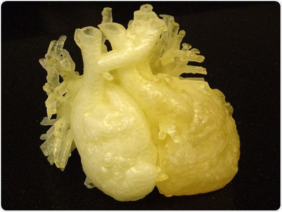
Mallott’s 3D printed heart model was used in the preoperative process to study his unique heart complexities
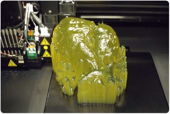
Dr. Ryan Moore and Dr. Michael Taylor from Cincinnati Children’s Heart Institute used Damon’s CT scan to generate a 3D reconstruction of his heart and convert it to a 3D printable file. Working closely with the CEAS Digital Fabrication Lab manager Sam Antoline, and his team of undergraduate engineering students, the 3D printed heart was fabricated with PolyJet-based 3D printing technology on the Objet260 Connex2 3D Printer from Stratasys using semi-translucent ABS and rubber-like TangoPlus materials. Antoline’s team was able to simulate characteristics of a human heart, experimenting with material-rigidity values (Shore A values) to create a model for surgeons and other medical personnel to use for pre-surgery planning.
31-year-old Damon Mallott was born with a complex congenital heart lesion for which he underwent creation of a single pumping chamber for his heart after multiple procedures as a child. The surgeries had proven to be a success, until recently when he started to develop severe heart-rhythm irregularities and a large blood clot in his heart. In order to improve his condition, he would need to undergo a challenging and rarely performed surgical procedure by Dr. David Morales, chief of cardiothoracic surgery. With the growing experience of 3D printing of congenital heart lesions at Cincinnati Children’s Hospital, Damon’s case provided the perfect opportunity to utilize a life-size scale model of his heart to stage his procedure.
This recent case is just one of over 20 successful heart surgeries performed as a result of surgical preparations with 3D printed heart models created through the collaboration of Cincinnati Children’s Hospital Heart Institute and the University of Cincinnati CEAS digital fabrication laboratory. Simulating surgery with a replica of a patient’s heart provides surgeons and their staff with an opportunity to assess complications and develop the best possible surgical plan for complex and risky operations. This type of pre-planning has an enormous impact on patient care and recovery. Additionally, the models serve as a source for educational tools—both for surgical training and educating patients and their families - about medical conditions and possible complications.

Patient heart model being produced on the Objet260 Connex2 3D Printer from Stratasys
The university’s CEAS Digital Fabrication Laboratory will continue to provide services for the continuing research at the Cincinnati Children’s Heart Institute. Working with 3D printing technology since 2006, the lab team has built various prototypes for programs ranging from the CEAS Medical Device Innovation and Entrepreneurship Program and aerospace research to university studies and combustion research. Antoline said a curriculum is likely to be developed in the near future, which would provide technical training to undergraduate students who are interested in learning more about 3D printing.
“We’re seeing a real demand for 3D printing technology at the university,” said Sam Antoline. “We’re producing prototypes and models for undergraduate capstones and university research across various departments. The detail we can achieve with this technology, and the ability to print with digital composite materials will definitely have a role within the university.”