Mar 24 2017
Oxford Nanoimaging Limited manufacture and sell custom microscopes offering super-resolution and single-molecule capabilities to research users. The multidisciplinary bioimaging unit, Micron Oxford, are using the Nanoimager instrument to advance their cellular imaging techniques for both their facilities and research programs.
Micron Oxford is a collaborative, multidisciplinary bioimaging unit working with biomedical researchers in the Oxford area and beyond, to apply advanced light microscopy imaging techniques to address key biological questions. Founded in 2010, Micron is supported by Wellcome Trust strategic funding. It has the remit to push the boundaries of imaging in light microscopy and to provide to its user community cutting edge, versatile tools.

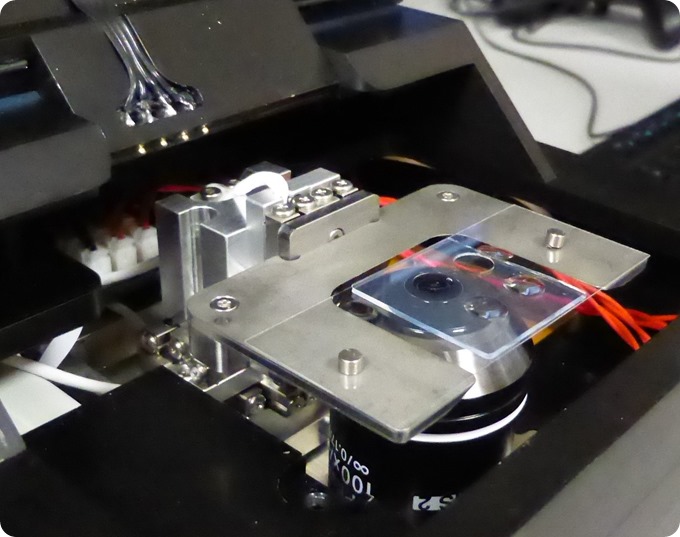
The sample chamber of the Nanoimager
Facility Manager, Andrew Jefferson, takes up the story. “We operate in a multidisciplinary scientific environment. Though based in the Department of Biochemistry, our users come from areas and departments across the scientific spectrum; from biology to chemistry, physics and photonics. As part of our remit at Micron, we always look to provide our users with the latest ground-breaking technologies. We have invested in super-resolution microscopy but not had an instrument that was accessible to all levels of user. That was until we discovered the Nanoimager from Oxford Nanoimaging (ONI).”

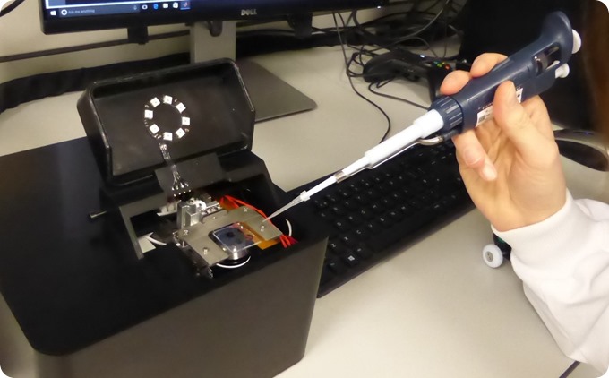
Pipetting a sample into the Nanoimager
The Nanoimager is a very small unit with a footprint of just 21 cm x 21 cm. It requires no special infrastructure such a vibration isolation tables, thermally controlled rooms or laser safety equipment. It has been designed at every point within its construction to compensate for both acoustic and vibration issues as well as fluctuations in temperature which are the nemesis of super-resolution techniques.
Loading a sample is as simple as pipetting onto a standard microscope slide. Then, very intuitive software guides the user through the selected imaging mode, first to capture raw data and to then process this to reveal ultra-high resolution, single molecule localised images. One of the initial projects is from the Bacterial Chromosome Dynamics group of Professor David Sherratt. Research in the Sherratt group is aimed at understanding how DNA replication, recombination and chromosome segregation shape bacterial chromosome organization in the context of the living cell.
PhD student, Florence Wagner, is using the Nanoimager for in vitro studies of structural maintenance of chromosomes (SMC) complexes. SMC proteins are present in all three domains of life and share a distinctive ring-shaped architecture that is believed to allow them to entrap and possibly reshape DNA molecules. Here she is reconstituting the DNA loading and unloading by SMC complexes purified from Escherichia coli bacteria cells to understand how these proteins interact with the chromosome and how this interaction relates to their biological function.

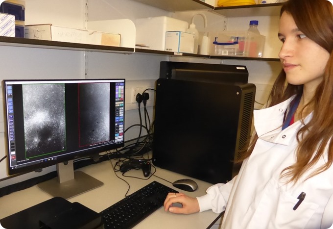
Florence Wagner of Micron Oxford working with the ONI Nanoimager to study structural maintenance of chromosomes (SMC) complexes at the single molecule level
In these experiments, both the proteins and the DNA are labelled with fluorescent dyes in order to image them in real time and characterise the nature and requirements of their association. Observing single SMC complexes as they act on the DNA will allow their function to be understood, as well the manner in which they maintain chromosomal architecture. Understanding how defects influence the function of these proteins is expected to provide insight into the processes associated with a number of diseases and syndromes.
Learn more about the possibilities of benchtop super-resolution and single molecule fluorescence microscopy and its applications at the Oxford Nanoimaging website: www.oxfordni.com.