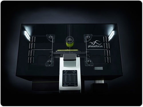
Phasefocus provides a range of products and services based on its proprietary Ptychographic Quantitative Phase Imaging (QPI) technology to provide a wide range of analytical applications including label-free cell imaging and quantitative electron phase microscopy. Here, they report on a poster presented at ASCB that shows the power of Livecyte for individual cell segmentation.

The Phasefocus Livecyte Cell Analysis System
Professor Norman Maitland's team from the Cancer Research Unit at the University of York presented a poster that showed the potential of Phasefocus’ Livecyte label-free cell analysis system as a rapid characterization tool of cell cultures derived from patient tissue.
“Characterization of the subpopulations of heterogeneous primary prostate epithelial cell cultures derived from patient tissue” sets out to address the issue that because traditional cell lines do not represent tumor heterogeneity or patient variability, there is a need for a better model to carry out pre-clinical testing in order to give greater chance of success in clinical trials. To achieve this, the use of primary cell cultures derived from patient tumors is becoming more desirable and more common. This is because primary cell cultures represent intra- and inter-patient heterogeneity. However, as the body contains a heterogeneous mixture of cells, to develop a successful treatment, it is necessary to target all cell types, and so combination treatments are likely to be more successful than monotherapies. The poster demonstrates the use of ptychography, a label free QPI (Quantitative Phase Imaging) technique, to characterize primary prostate epithelial cultures derived from patient tumor tissue. QPI enables the phase information of light to be retrieved, allowing the user to obtain multi-parametric information relating to the intrinsic properties and behavior of each individual cell.
The experiments showed that using the Livecyte system, it was possible to characterize the differences in cell behavior between a primary prostate cancer cell population and that of conventional cell lines. Comparisons were made of separate sub-populations from primary prostate cell cultures using immunofluorescence and flow cytometry. The sub populations were characterized by the identification of morphological and kinetic phenotypes at an individual cell level. In contrast to the immunofluorescence and flow cytometry approach, the cell population was not destroyed to enable the characterization, and as such the population can be continued to be imaged and further analyzed. Using the Phasefocus Livecyte system, clear phenotypic differences were observed between different populations. Since Maitland and his team were able to identify key differences between the two known subpopulations in primary prostate cultures using QPI, they went on and tested out those parameters on a heterogeneous culture of primary prostate cells and were successfully able to identify the two subpopulations within the mixed culture. This shows that cells can be identified by a unique ptychographic signature.
Looking to the future, full characterization of all types of patient-derived tumor cells, alongside analysis of their response to current and novel drugs will allow assessment of detailed biological effects of the drugs tested as well as identification of resistant cells. This could lead to patient cells becoming part of the drug development pipeline, which will ultimately result in targeted and patient stratified therapies that take into account intra- and inter-tumor heterogeneity.