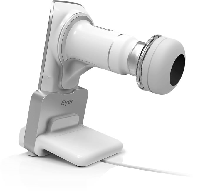
A portable device attached to a smartphone can help capture precise images of the retina to diagnose eye disease. The new method by a startup company, Phelcom Technologies, is a lower cost tool that can help doctors diagnose remotely, through telemedicine.

An app operating the optical device sends images of the retina over the internet to Eyer Cloud, which stores and manages patient files. Image Credit: Phelcom Technologies
The device, called Eyer, is designed to light up and visualize the retina. It is connected to a smartphone’s camera. An application sends the captured images over the internet via the Eyer Cloud, which stores the patient files. The ophthalmologist can view the images remotely through the cloud by accessing the patient files.
Due to lack of internet connection in some areas across the globe, the images are kept in the smartphone app and sent to the cloud when an internet connection, either Wi-Fi or 4G network is available.

Phelcom Eyer
Help diagnose retinal eye diseases
The new device can help diagnose eye diseases, particularly those affecting the retina. The retina is the light-sensing tissue found in the back of the eye. One of its roles is to relay images to the brain.
Back-of-the-eye or fundus eye diseases include conditions such as Stargardt’s disease, macular degeneration, retinal tear, diabetic retinopathy and retinal detachment, among others. Stargardt’s disease refer to a group of inherited diseases that affects the light-sensitive cells in the retina to deteriorate. Macular degeneration is a condition where the center of the retina deteriorates, leading to blurred central vision or a blind spot. Retinal tear happens when the vitreous, or gel-like substance in the center of the eye shrinks, pulling the thin layer of tissue lining in the retina, causing a tissue break. Diabetic retinopathy can cause blindness in people with diabetes. It happens when the blood sugar shoots up, causing damage to the blood vessels supplying the retina.
The Eyer Project
In March, Phelcom established its São Carlos factory after getting a certification from the National Health Surveillance Agency (ANVISA) and the National Institute of Metrology, Quality and Technology (INMETRO).
The factory manufactures 30 units a month but is projected to reach about 100 units by the end of 2019. The device is priced at $5,000, accompanied by a high-quality smartphone. The device is way cheaper than conventional equipment. The currently-used ophthalmoscope device has to be connected to a computer to capture images and it costs around $30,000.
“We invested significantly in optics and in design. One challenge was producing a portable version of a device that is typically very large. Another was enabling nonmydriatic operation so that high-quality images of the retina can be captured without the need for pupil dilation,” José Augusto Stuchi, Phelcom CEO, said in a statement.
The Eyer Cloud
The company has also developed the Eyer Cloud, an innovation designed to store and manage all the data, including patient information and retinal images, acquired in the diagnostic test. The software works by organizing data, so doctors can remotely access for diagnosis.
In contrast, current equipment used today needs to be attached to a computer. The Eyer is portable, easier to use, and more accessible. To set up the Eyer and its cloud, the user should create an account, where the images can be automatically saved.
The researchers assured that all data are kept private and confidential. Also, they worked on making the transmitting of images from the device to the cloud faster, so doctors can access the images regardless of the device’s location.
The device uses telemedicine, since a licensed or trained technician uses the device to capture the images and sends the photos to the cloud. Meanwhile, an ophthalmologist can visualize and analyze the photos in another location. The process makes diagnosing faster and more convenient.
At present, the company plans to establish a partnership with ophthalmologists to develop a part of the system responsible for reporting. For doctors, the planned payment is by a monthly subscription and each report will roughly cost about $5 to $10.
Artificial intelligence
Aside from the cloud, the team is working on artificial intelligence to help in finding patterns, detecting diabetic retinopathy. The medical reports will be placed in a database, where the computer uses AI to find patterns linked to retinal diseases.
A software, approved by the U.S. Food and Drug Administration (FDA), called IDx-DR, utilizes AI to examine eye images captured by a retinal camera. These images will be analyzed to detect diabetic retinopathy, potentially diagnosing the condition early to prevent blindness.
The company has more than 10,000 images of the retina and projects about 50,000 patients next year. They plan to have the largest database across the globe.