Scientists have come up with a very rapid 3D printing technique for producing the most complex tissue forms in hydrogels which are loaded with stem cells, and then generating the growth of blood vessels in the resulting tissue mass.
This revolutionary optical technique could transform the way tissue engineering is done, allowing the production of all kinds of tissues and even organs. It could be possible to design tissues on which to test new drugs, repair damaged parts of the body, and even replace organs made to order, to fit each individual while being fully functional.

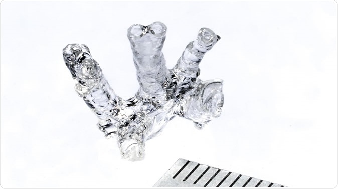
Hollow mouse pulmonary artery model © Alain Herzog / 2019 EPFL
Living tissues are extremely functional chiefly because of their highly intricate design. Shape is closely linked to function, in the body. Free-form shapes are essential for cancellous bone and within menisci in the knee, for instance. The irregular multidirectional nature of bone trabeculae in the former, and of carefully aligned fibers in the latter, is essential to their function.
This is where conventional tissue synthesis methods like stereolithography and extrusion-based tissue printing have run into a number of problems. For one, they produce tissues layer by layer, and produce arrays of repeated structures like single cells, voxels or groups of cells. This limits the type of internal design, since replicating porous living tissues, for instance, requires the use of struts.
Bioprinting complex living tissue in just a few seconds
Secondly, the long time taken to print a useful amount of tissue means that cells are exposed to non-optimal environments for longer periods, which reduces the number of functional cells that can be eventually obtained.
In the current study, researchers put their heads together to come up with a completely different technique that takes just a minute or so to produce the right 3D form inside the hydrogel, into which stem cells can grow to produce a tissue model with exactly the right architecture. All that remains is to add endothelial cells to produce the growth of blood vessels into this mass. This is because the whole structure is printed rather than one layer at a time.
The technique called volumetric bioprinting was developed at the Laboratory of Applied Photonics Devices (LAPD), in EPFL's School of Engineering, in collaboration with other scientists at Utrecht University.
The study appears in the journal Advanced Materials.
How is volumetric bioprinting done?
Volumetric bioprinting makes use of laser projection into the interior of a rotating cylinder loaded with the hydrogel which is filled with stem cells. These cells are capable of differentiating into any type of cell, depending on the environmental and chemical cues. Optical light is shaped into a series of 2D patterns which are beamed into the spinning light-reactive polymer or hydrogel to achieve a cumulative 3D distribution of the total dose required.
The laser energy thus builds up to a focus at specific points. These points on which the laser energy builds up to a threshold dose then undergo polymerization to become solid. This therefore forms an intricate 3D shape suspended within the hydrogel, leaving 85% of the stem cells intact. Finally the endothelial cells are loaded into the mass to grow new blood vessels.
Using carefully measured light intensities and the right amount of photoinitiator, a compound which triggers the polymerization, they found that within seconds, a complex structure could take form.
How is it better?
The advantages include the ability to scale up the size of the model without increasing the build time greatly. The surface of the model appears to be free of artifacts, reproducing the smoothness of the digital model faithfully. Thirdly, there is no need to add support material which must later be sacrificed. This allows free-floating designs to be executed. Fourthly, cells preserve their viability, and continue to function normally over time. Finally, there is no damage due to shearing stress or other damage, ensuring their function remains intact. The early thigh tissue models suggest that the bioprinted tissue can achieve important cellular functions.
The tissue can be as large as several centimeters using this process, which allows them to construct tissues of a useful size. To demonstrate the feasibility of their approach, they have already produced a structure resembling one of the human heart valves, , and part of the thighbone with a complicated shape, with buds of endothelial tissue ready to grow into the interior. A meniscus that incorporates the cartilage forming cells has also been formed, and these cells produce new matrix just as in the body, allowing the graft to become mature in vitro. Interlocking structures have also been built using the same procedure.
The volumetric bioprinting is amazingly fast, and allows the build to be designed with greater freedom. This is in contrast to the layered build of conventional bioprinting which takes a long time.
Researcher Paul Delrot says, “The characteristics of human tissue depend to a large extent on a highly sophisticated extracellular structure, and the ability to replicate this complexity could lead to a number of real clinical applications.” The capability of churning out artificial organs and tissues at heart-stopping speed will greatly facilitate drug development and testing in the early stages, avoid animal testing, and contribute to immense cost and time savings, not to mention the ethical benefits of phasing out animal tests. This justifies the encomiums of LAPD head Christophe Moser: “We believe that our method is inherently scalable towards mass fabrication and could be used to produce a wide range of cellular tissue models, not to mention medical devices and personalized implants.”
And they plan to do just that, creating a partner company to market their innovative technique.
Source:
actu.epfl.ch. (2019). Bioprinting complex living tissue in just a few seconds. https://actu.epfl.ch/news/bioprinting-complex-living-tissue-in-just-a-few-se/
Journal reference:
Volumetric Bioprinting of Complex Living‐Tissue Constructs within Seconds. Paulina Nuñez Bernal, Paul Delrot, Damien Loterie, Yang Li, Jos Malda, Christophe Moser, & Riccardo Levato. Advanced Materials. 19 August 2019. https://doi.org/10.1002/adma.201904209. https://onlinelibrary.wiley.com/doi/full/10.1002/adma.201904209