Electrodes are used to pick up electrical signals. Neurons however are tiny units of the nervous system and it is difficult to create an electrode small enough to record all the workings of a neurons.
Researchers from Harvard University have overcome this obstacle by creating an electronic chip that could be implanted within these networks and could thus perform highly sensitive intracellular recordings of the neurons individually. This could be a boon for neuronal research believes the team as it could help provide an in-depth knowledge of the neuronal connections and synaptic workings.
The team wrote, “Current electrophysiological or optical techniques cannot reliably perform simultaneous intracellular recordings from more than a few tens of neurons.” The study results with the newly developed chip were published in the latest issue of the Nature Biomedical Engineering. The study is titled, “A nanoelectrode array for obtaining intracellular recordings from thousands of connected neurons.”

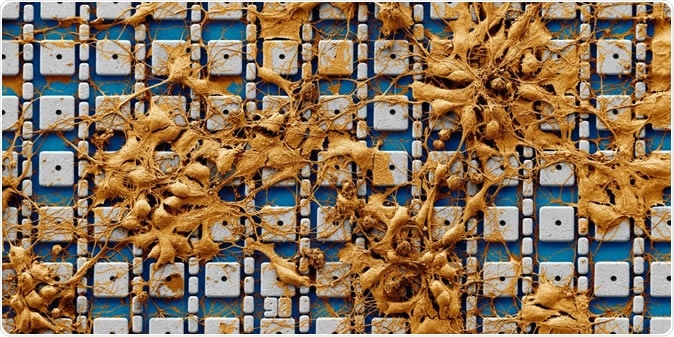
False colored scanning electron microscope image of neurons cultured on top of the electrode array. Actual recording experiments are performed with much higher neuron densities containing three to six cell layers covering the entire electrode array. Image Credit: Harvard SEAS
Hongkun Park, Mark Hyman Jr. Professor of Chemistry and Professor of Physics, and one of the senior authors of the study explained, “Our combination of the sensitivity and parallelism can benefit fundamental and applied neurobiology alike, including functional connectome construction and high-throughput electrophysiological screening.” Donhee Ham, Gordon McKay Professor of Applied Physics and Electrical Engineering at the John A. Paulson School of Engineering and Applied Sciences (SEAS), another senior author added, “The mapping of the biological synaptic network enabled by this long sought-after parallelization of intracellular recording also can provide a new strategy for machine intelligence to build next-generation artificial neural network and neuromorphic processors.”

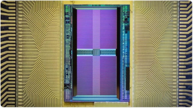
The electronic chip uses the same fabrication technology as computer microprocessors. (Image courtesy of Harvard SEAS)
The team explained that this chip is much like the microprocessors used in computers and the chip contains vertical nanometer scale electrodes that are arranged in a dense cluster. These are operated using a high-precision integrated circuit. Each of the nanoelectrodes are coated with platinum powder which makes their surfaces rough and this allows them to convey the signals. They wrote, “The array consists of 4,096 platinum-black electrodes with nanoscale roughness fabricated on top of a silicon chip that monolithically integrates 4,096 microscale amplifiers, configurable into pseudocurrent-clamp mode (for concurrent current injection and voltage recording) or into pseudovoltage-clamp mode (for concurrent voltage application and current recording).”

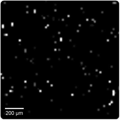
Intracellular recordings of neurons across a connected network. The videos are slowed 4× from real time. (Video courtesy of Harvard SEAS)
In order to test the neurons, the team cultured the neurons on the chip. The circuit is then programmed to send in a signal via the nanoelectrodes into the neurons. This opens up microscopic holes on the neuronal surface, they explained. This opens up access within the neurons, they added. These holes allow the nanoelectrodes to detect the voltage signals that travel medicated by the neurons. Jeffrey Abbott, a postdoctoral fellow in the Department of Chemistry and Chemical Biology and SEAS, and the first author, in a statement said, “In this way we combined the high sensitivity of intracellular recording and the parallelism of the modern electronic chip.”
For this study the team looked at over 1,700 rat neurons and over twenty minutes of neuronal recording, they found over 300 synaptic connections and found the interplay of the neurons in the network. Abbott explained, “We also used this high-throughput, high-precision chip to measure the effects of drugs on synaptic connections across the rat neuronal network, and now we are developing a wafer-scale system for high-throughput drug screening for neurological disorders such as schizophrenia, Parkinson's disease, autism, Alzheimer's disease, and addiction.” The authors wrote in conclusion, “This high-throughput intracellular-recording technology could benefit functional connectome mapping, electrophysiological screening and other functional interrogations of neuronal networks.”
On the team with the above wuthors were Tianyang Ye, Keith Krenek, Rona S. Gertner, Steven Ban, Youbin Kim, Ling Qin and Wenxuan Wu. This study was funded and supported by Samsung Advanced Institute of Technology, Catalyst Foundation, the US Army Research Office, the National Science Foundation, the National Institutes of Health, and the Gordon and Betty Moore Foundation.
Related study
Couple of years back researchers led by David Jackel published a related study called, “Combination of High-density Microelectrode Array and Patch Clamp Recordings to Enable Studies of Multisynaptic Integration,” published in the journal Scientific Reports.
The team wrote that they could record the electrical activity of the neurons from tens in an array including presynaptic neurons. The team used parallel mapping to look at the synapses. They used 11000 electrodes for their experiment for extracellular recording and stimulation and also used intracellular patch-clamp recording. With their set up the team had successfully identified the contributions of the individual presynaptic neurons and this helped them look at dendrite networking of the cortical neurons.
Journal reference:
Jeffrey Abbott, Tianyang Ye, Keith Krenek, Rona S. Gertner, Steven Ban, Youbin Kim, Ling Qin, Wenxuan Wu, Hongkun Park & Donhee Ham, A nanoelectrode array for obtaining intracellular recordings from thousands of connected neurons Nature Biomedical Engineering (2019), DOI: 10.1038/s41551-019-0455-7, https://www.nature.com/articles/s41551-019-0455-7