Scientists have found out a way to forecast the spread of brain atrophy, using prior research that shows how brain networks connect with each other to form a continuously linked pathway. The research published in the journal Neuron on October 14, 2019, was carried out in patients with frontotemporal dementia (FTD) and shores up earlier findings that patients with dementia lose brain cells to atrophy in a systematic pattern, that follows the synaptic connections between neurons in already well-defined networks. This tells us more about the progress of neuronal degeneration – where the nerve cell or pathway is at greatest risk for atrophy – as well as helping thereby to evolve better tools, in the foreseeable future, to help measure how efficiently new treatments can block this process.
Dementia is an umbrella term for any condition that is caused by the degeneration of brain cells resulting in altered behavior and difficulties in language use – the most well-known condition in this category being Alzheimer’s disease. FTD is the most common form of dementia to affect people below the age of 60 years. FTD affects different people in different ways, the variation in the effects being mediated by the route of spread of the atrophy through the brain in each case. This high degree of unpredictability makes it harder to specifically point to any one factor as the underlying biological reason for atrophy. It also makes it harder to design a clinical trial to see whether a new treatment is actually working in any way, or is superior to previous treatments.

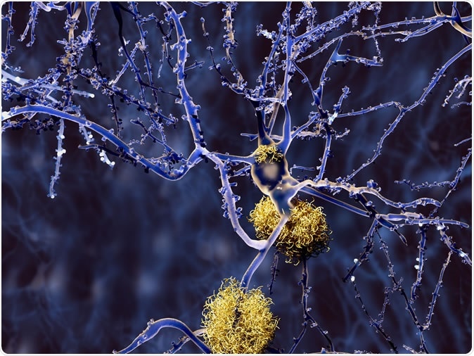
Alzheimer's disease: neuron with amyloid plaques. Image Credit: Juan Gaertner / Shutterstock
Mapping functionally connected neural networks
The first sign of change in this research field came from the current team’s senior researcher William Seeley, who showed how atrophy actually follows the pathway of an already existing brain network in use. A brain network is a community of neuronal clusters that are related to the same function, such as speaking, hearing, language processing and the like. These networks communicate and work together because of the connections shared by the neurons within them – the synapses. These clusters may be nearby or far apart from each other. Thus the progression of cell death in neurodegenerative conditions is not random, nor is it like a wave of degeneration spreading outwards in all directions from a central focus. Instead, it is like a telegraphic message spreading along a communication pathway, composed of the various physical devices that make up the circuit.
The study
The current study takes forward the same concept by showing that they can predict how brain atrophy will progress, in a patient with FTD, based on the maps of neural networks in the brain, derived from brain scans in healthy people. The researchers had already created a standardized map of 175 neural functional pathways in different parts of the brain, based on functional MRI scanning of 75 healthy people. Functional MRI uses vascular flow detected by MRI in various parts of the brain, to assess which part of the brain is functioning during a given activity.
The research included 42 patients with FTD who had altered behavior – a type called behavioral variant FTD – in the form of inappropriate social behavior; and 30 patients with FTD of the semantic variant that showed up as primary progressive aphasia – where the main symptom is the marked deterioration in the patient’s ability to use language.
On an initial baseline MRI scan, these patients were assessed to see how much of the brain had already degenerated. One year later, or so, a repeat MRI was taken to evaluate the progression of brain atrophy in this period.
Once the maps were in place, the researchers matched the maps from the FTD patients one by one to the standardized neural network maps based on best-fit – looking at which of the standardized networks matched the atrophy pattern in the affected individual’s baseline MRI. They then arbitrarily assigned the position of ‘origin of atrophy’ to the center of the best-matching network. The underlying assumption was that neuronal atrophy begins at a especially high-risk location in the network, and then spreads to other neural circuits through the pre-existing synapses. Thus the ‘hub’ of any anatomically connected network is the logical place for the initial atrophy to begin.
Using these standardized maps, they predicted the most likely route of spread of atrophy over the following year. They then tested their predictions against the follow-up set of MRI scans done a year later. They also compared the accuracy of these predictions against those which were done independently of functional connectivity within the affected network.
The findings
The researchers found that they needed to include two steps, in particular, to make the most accurate prediction of whether any given brain region would be involved in the atrophy over the following year after the baseline MRI scan.
The first is the “shortest path to the epicenter,” and is taken as the number of synapses crossed by the atrophic process from the supposed epicenter of the degeneration, up to the region where a prediction is sought. This corresponds to how many synapses link the two regions via the neural network. In other words, the more synapses there are, the slower the speed of progression to this region.
The second step to be factored in is the “nodal hazard”, or the number of regions within the connectivity range that are already showing significant degeneration. This is important because the more regions that are already affected, the greater the chance of the target region being affected as well.
As researcher Jesse Brown says in his analogy, “It's like with an infectious disease, where your chances of becoming infected can be predicted by how many degrees of separation you have from 'Patient Zero' but also by how many people in your immediate social network are already sick.”
The scientists saw a lot of hope in their method, but also pointed out that a lot of work is still awaiting them. They need to make their predictions much more precise, such as by using individualized maps to show the neural network connectivity for each patient rather than a standardized map, or by classifying maps by subtype and developing more narrow models for each type.
Importance of this study
The present study tells scientists more about how brain atrophy spreads in FTD via the biological underpinnings of the disease progression. This will, of course, eventually trigger the development of drugs to avoid or slow down such spread. In an apt comment, Brown sums up: “Just like epidemiologists rely on models of how infectious diseases spread to develop interventions targeted to key hubs or choke points. Neurologists need to understand the underlying biological mechanisms of neurodegeneration to develop ways of slowing or halting the spread of the disease.”
Moreover, it can help to build tools that will tell researchers how well a particular therapy works – is it actually changing the predicted route or speed or distance of progression over the network?
As the atrophic process destroys increasing numbers of brain neurons and areas, the devastating changes in the patient’s personality and behavior will continue to alter. The heavy load this imposes on the loved ones and caregivers can be somewhat alleviated by bringing down the degree of unpredictability, since this model could help them foresee some of the likely changes ahead with advanced disease. This could also help them take proper decisions and to prepare for such changes.
William Seeley says, “We are excited about this result because it represents an important first step towards a more precision medicine type of approach to predicting progression and measuring treatment effects in neurodegenerative disease.”
Journal reference:
Patient-Tailored, Connectivity-Based Forecasts of Spreading Brain Atrophy Brown, Jesse A. et al., Neuron, DOI:https://doi.org/10.1016/j.neuron.2019.08.037, https://www.cell.com/neuron/fulltext/S0896-6273(19)30743-3