A new compound has been invented to bind to liver cells that are in the early stages of damage, thus making it easier to pick them up using MRI (magnetic resonance imaging).
The use of this compound prevented the need to do the invasive procedure called liver biopsy to arrive at an assessment of the stage of liver damage or its severity – a marked advance, since biopsy (removal of a small section of tissue for microscopic examination) is the gold standard for the diagnosis and staging of chronic liver disease.

Image Credit: VILevi / Shutterstock
Chronic liver disease
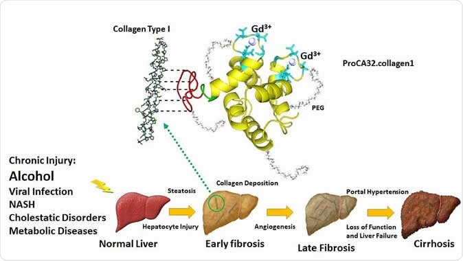
Within just 5 years (2010 to 2015), chronic liver disease led to 31% more deaths in the age group 45-64 years, in the US. Chronic liver damage may be due to non-alcoholic fatty liver disease that affects up to 40% of the US population, as a result of obesity, viral hepatitis, or autoimmune disease; or alcoholic liver damage due to drinking too much. Toxins primarily induce liver damage and finally death of the cells, followed by an attempt to heal via fibrous scarring. This disrupts the delicately and accurately placed arrangement of liver and blood vessel tissues, reducing liver function and possibly causing more damage.
Though this is a slow process, taking years to manifest, it is usually far advanced and the consequences of failing to detect or treat it early are very severe. Current tests cannot pick up liver fibrosis, at its earliest stage, with sufficient accuracy. As a result, says Yang, “Most people do not believe they have liver fibrosis and don't want to change their lifestyle. They continue and at some point develop later-stage fibrosis which can become severe cirrhosis and a large portion become liver cancer.”
ProCA32.collagen1 – numerous benefits
All that could now change with the new contrast agent. MRI is already highly preferred in imaging because it avoids ionizing radiation, penetrates tissue deeply to reduce the chance of missing a lesion within the liver, has excellent resolution and covers the whole liver. A contrast dye for MRI scanning is meant to make selected body structures more easily visible.

Novel collagen-targeted protein MRI contrast agent enables non-invasive detection of early stage of liver fibrosis by MRI. Jenny J. Yang, Ph.D., Georgia State University
ProCA32.collagen1 is designed to bind very well to collagen I, a fibrous protein that is increased in fibrotic liver cells. This binding renders them visible because of the bound gadolinium, a heavy metal which shows up well on MRI.
In this way, the black-and-white imaging of ProCA32.collagen1 distinguishes previously invisible fibrotic tissue from other healthy liver tissue in the background. Not only so, it also avoids the possibility of missing a single or a few fibrotic regions scattered in a sea of healthy tissue, as may occur with traditional liver biopsy. This is one primary reason why liver biopsy is notorious for high error rates, up to 30% to 40%, even at advanced stages of fibrosis.
Animal tests have shown that it binds well to collagen fibers within liver cells affected by early liver fibrosis, both alcoholic and non-alcoholic. It is able to detect early fibrosis twice as reliably as traditional contrast agents. It also differentiates early non-alcoholic liver fibrosis from late fibrosis and from normal liver tissue.
It can help correctly stage and assess the severity of fibrosis in this condition. Moreover, it can help monitor treatment of liver fibrosis to assess the efficacy of the therapy.
Another advantage is its ability to pick up metastatic microtumors a hundred times smaller than the smallest diameter visible by any currently used contrast agent. This was published in the journal Biomaterials. Thus a mass sized just 0.1mm to 0.2mm may be seen on MRI with the use of ProCA32.collagen1.
A final conclusive advantage is the very low dosage as well as its 108 to 1016 fold preference for gadolinium compared to other ions more commonly found in the body – like zinc or calcium. This means the chance of releasing gadolinium in the body is minimal – which means a greatly lowered risk of toxicity with gadolinium.
This protein-based agent can also do color imaging, to pick up different features in different colors. This will increase both sensitivity and accuracy, say the researchers. They now plan to submit the patented contrast agent to the US Food and Drug Administration (FDA) for approval so they can hold clinical trials with it.
Implications
Researcher Jenny Yang says it’s “a revolutionary change”, and will “help doctors monitor treatment before it is irreversible and help pharmaceutical companies to select the right patients for clinical trials or identify subjects for drug discovery.”
The report concludes: “The contrast agent is expected to overcome the major clinical barriers in early diagnosis, noninvasive detection and staging of chronic liver diseases, and have strong translational potential for monitoring treatment efficacy.”
Source:
Journal reference:
M Salarian, RC Turaga, S Xue, M Nezafati, K Hekmatyar, J Qiao, Y Zhang, S Tan, OY Ibhagui, Y Hai, J Li, R Mukkavilli, M Sharma, P Mittal, X Min, S Keilholz, L Yu, G Qin, AB Farris, Z-R Liu, and JJ Yang. Early detection and staging of chronic liver diseases with a protein MRI contrast agent. Nature Communications 10, Article number: 4777 (Oct. 29, 2019). DOI: 10.1038/s41467-019-11984-2. https://www.nature.com/articles/s41467-019-11984-2