Pulmonary embolism is a condition where a clot forms and blocks the arteries of the lungs and this restricts the blood flow to the lungs and lowers the amount of oxygen reaching the different organs. Statistics from the National Institutes of Health show that pulmonary embolism leads to nearly 200,000 to 300,000 deaths in the United States annually putting it at number three in cardiovascular diseases right after heart attack and stroke. Pulmonary embolism can be life threatening and is usually treated by clot busting medicine or manoeuvres or surgeries.

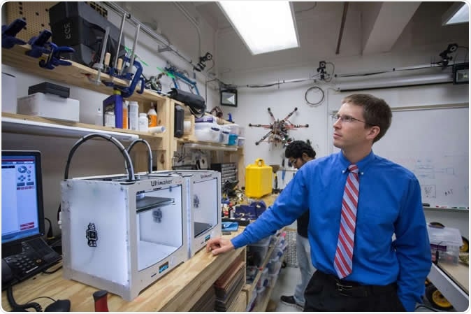
Aaron Becker, assistant professor of electrical and computer engineering at the University of Houston, is leading a project to use swarms of tiny robots to break up blood clots. Credit: University of Houston
Researchers from University of Houston's Robotic Swarm Control Laboratory and Houston Methodist Hospital, now have come up with tiny robots that could be sent to the site of the clot within the body. These robots could break down the clots and open up the blockage say the researchers. Aaron Becker, principal investigator of the study and Assistant professor of Electrical and Computer engineering at the UH Cullen College of Engineering, said in a statement, “Our lab is at the forefront of developing magnetically controlled tiny robots, and we have a strong collaborative relationship with the Methodist Research Institute. This research is motivated by clinical problems [physicians] deal with.” This project was titled, “Wireless magnetic millibot blood clot removal and navigation in 3D-printed patient-specific phantoms using Echocardiography,” and was funded by the National Science Foundation with a grant amount of $752,871.

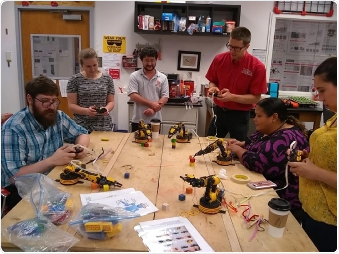
UH professor Aaron Becker (red shirt) and Julien Leclerc (next to him, gray UH shirt), a UH Cullen College research associate visit with Houston-area teachers and an undergraduate REU participant in the Robotic Swarm Control Laboratory. Credit: University of Houston
For this project the team would use tiny robots of the size 6 mm by 2.5 mm shaped like a corkscrew. Each of these robots have a small rotating swimmer with a magnet. These robots would be guided within the body to the site of the clot using magnetic waves. Ultrasound imaging would be used to locate the robots within the body and a set of electromagnets would be used to control their movements. Within the arteries they would help break up the clots and open up the blockages. Julien Leclerc, a research associate at the UH Cullen College of Engineering, explained, “It reminded me of an episode of 'The Magic School Bus,' where the vehicle is miniaturized and navigates inside Ralphie, a student, to find the cause of his sickness. I immediately liked the concept of using magnetic fields to control tetherless miniature robots inside a patient.”

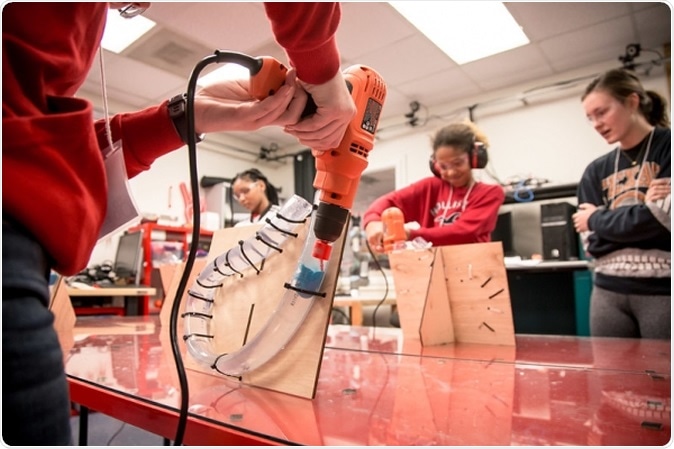
GRADE camp students visit Dr. Aaron Becker and the UH Robotic Swarm Control Lab. The blue mass represents the blood clot the participants are trying to break up. Credit: University of Houston
Others working on the team included Dr. Dipan Shah, chief of the Division of Cardiovascular Imaging, and Mohamad Ghosn, a research scientist with the DeBakey Cardiac and Cardiovascular Center. The team explains that pulmonary embolism could be just one of the uses of these miniature magnetically-controlled robots. There may be other uses for these including complex brain surgeries and eye surgeries and clot removal from tiny blood vessels.
Leclerc said, “Using non-invasive miniature magnetic agents could improve patient comfort, reduce the risk of infection and ultimately decrease the cost of medical treatments. My goal is to quickly bring this technology into the clinical realm and allow patients to benefit from this treatment method as soon as possible.”
Pulmonary embolism
Pulmonary embolism is managed by a multidisciplinary Pulmonary Embolism Response Team (PERT). Diagnosis is made initially by testing for D-dimer followed by a CT angiography imaging study. Echocardiography and/or portable V/Q scan, duplex ultrasonography are also suggested. In addition vital parameters including systolic blood pressure, heart rate, respiratory rate, respiratory rate, oxygen requirement etc. need to be recorded.
Treatment of pulmonary embolism involves use of anticoagulant medication. Researchers have said that treatment needs to be started based on suspicion of pulmonary embolism alone even before confirmed diagnosis is obtained because of the life-threatening nature of the condition. To break down the clot thrombolytics are used as injections. Again treatment needs to start as soon as possible and immediately in cases of cardiac arrest seen in suspected pulmonary embolism patients. The clots could also be broken by inserting a catheter in a procedure called Catheter Directed thrombolysis (CDL) or Catheter Directed Embolectomy. These are necessary for patients with intermediate to high risk of pulmonary embolism say researchers. These are opted as first line of action is systemic thrombolysis cannot be chosen. The next step is Surgical Embolectomy (SPE) which involves removal or breakage of the clot by open surgery. This is tried only when systemic thrombolysis and catheter directed thrombolysis has failed or the patient is at a very high risk. Surgery is also a choice if the thrombus lies largely in the right heart and is unwieldy and big in size.
In addition to the interventions hemodynamic support is provided and filters are placed within the large vein Inferior Vena Cava to prevent movement of the clot. The patients after discharge need to be followed up within 2 weeks or three months or earlier with symptoms.