.jpg)
Credit: LullaBEE / Shutterstock
In a pregnancy ultrasound, high-frequency sound waves (too high for the human ear to hear) are beamed at the uterus to reflect back in varying patterns, depending on the distribution of the tissues of the uterus and the baby. The reflected waves are collected by a receiver and analyzed by a software program to depict the image of the fetus, its heartbeat, its movements and the surrounding uterus. This technique is used to pick up a number of complications that can occur in pregnancy.
Attenuation coefficient
As ultrasound waves travel through tissue, they lose amplitude (size) and intensity (energy), and this change is called attenuation. If the distance through which they travel is fixed, waves at higher frequency will be more affected, or will weaken more, than those at lower frequency. For this reason, lower frequencies are used to examine areas which are deeper in the body. This helps provide an image of these deeper structures, but the resolution will be less clear.
Attenuation occurs because the energy of the ultrasound wave interacts with the tissue, in various ways including absorption, scattering, reflection, and interference. The energy of the wave is primarily converted into heat energy or other forms of energy, by absorption, and such conversion is the chief reason for attenuation. Scattering is another contributor. The extent to which a given medium produces attenuation of the ultrasound wave at a specific frequency is represented by the attenuation coefficient. The highest attenuation coefficient is for aerated lung tissue, which allows virtually no ultrasound waves to pass through. The lowest is for blood and water, for which attenuation is negligible.
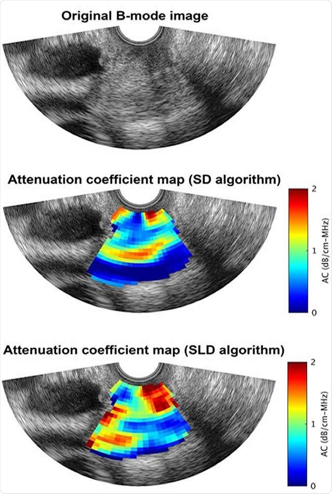
The team of researchers focus on the attenuation coefficient obtained by ultrasound imaging of the cervix, which helps them evaluate any cervical changes in pregnancy and in the pre-labor phase. They hope this will translate into an early warning system of impending preterm labor, before symptoms like pain, uterine contractions or cervical dilation occur. Two techniques were used to find the attenuation coefficient: the spectral difference and the spectral log difference.

An ultrasound B-mode image of the human cervix superimposed with a) anatomy labels, b) an attenuation coefficient map obtained using the spectral difference (SD) algorithm, and c) an attenuation coefficient map obtained using the spectral log difference (SLD) algorithm. Image Credit: Aiguo Han
The cervix
The female cervix is a firm cylindrical structure that guards the uterine opening into the vagina, about 25 mm long and 20-25 mm wide. It has a fibrous stroma covered on the internal aspect with one layer of columnar epithelial cells that secrete mucus. The stroma is formed primarily of collagen, which accounts for the firm feel of the cervix. This forms about 80% of the stroma, with its ground substance. The collagen is oriented both circularly around the cervical canal in the center, that communicates with the uterus at one end and the vagina at the other, and longitudinally to prevent premature shortening of the cervix.
As pregnancy proceeds, the cervical canal widens a little, as does the stroma, perhaps because the collagen is broken down and the collagen network weakens as a result. The volume of the cervix increases because of these changes, and this helps keep the cervix closed. As the third trimester begins and progresses, the cervix begins to shorten. With the near approach of labor, remodeling begins, with collagen realignment, and altered organization. This causes the cervix to soften, ripen, dilate and finally to regain its prepregnant characteristics following childbirth.
The study
The scientists say they built their study upon the biological basis of cervical changes that occur during pregnancy and before childbirth, such as remodeling of collagen. These changes are sensitively recorded by ultrasound, since the attenuation of the cervical collagen fibers is high in pregnancy because of the tight packing pattern. Researcher Barbara L. McFarlin says, “In preparation for labor and birth, there is increased collagen disorganization, increased water content and inflammation of the cervix, with low ultrasonic attenuation.”
The study involved compiling data on the attenuation coefficient values derived from the use of both techniques. They then compared the data from each technique for sensitivity and accuracy of prediction with respect to changes in the cervix.
Implications and follow up
The researchers did not compare the data with the final clinical outcome in this study. Aiguo Han, who also worked on the project, says more study will be needed to assess the performance of both techniques to find out which correlates well with the clinical picture so that their diagnostic value can be evaluated. This could eventually help doctors predict preterm birth and take preventive measures to delay birth if possible, or if required.
Journal reference:
Comparison of two spectral-based techniques for estimating the attenuation coefficient from human cervix, Ziyang Xu, Aiguo Han, Douglas G. Simpson, W. D. O'Brien, Jr., Barbara L. McFarlin, The Journal of the Acoustical Society of America 146:4, 2864-2865, https://asa.scitation.org/doi/abs/10.1121/1.5136939