A new form of magnetic resonance imaging (MRI) that makes cancerous tissue glow in medical images could help doctors more accurately detect and track the progression of cancer over time.

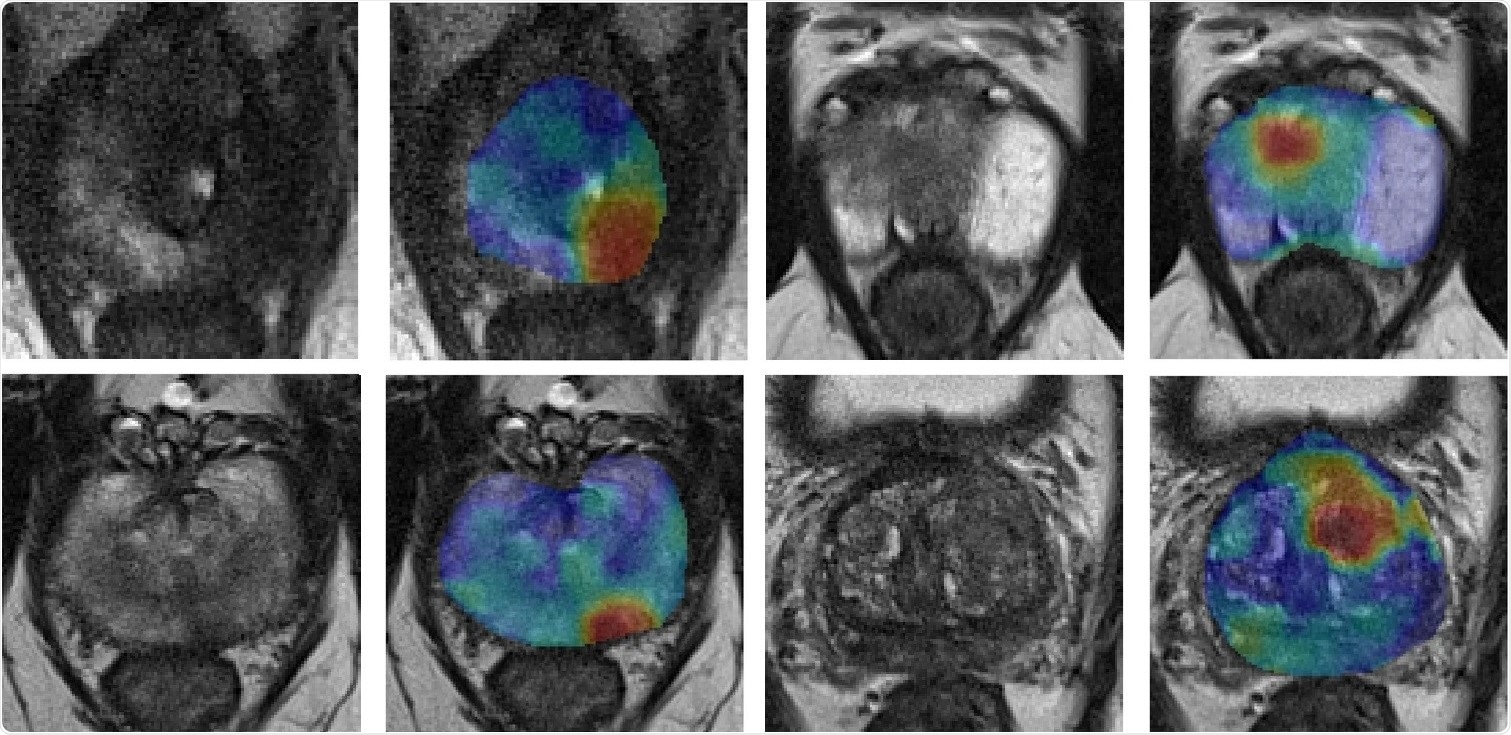
The images on the left are standard MRI of a prostate gland. The same prostrate is shown to the right of each image with a cancerous tumor highlighted in red, using new MRI technology called synthetic correlated diffusion imaging. Image Credit: University of Waterloo
The innovation, developed by researchers at the University of Waterloo, creates images in which cancerous tissue appears to light up compared to healthy tissue, making it easier to see.
Our studies show this new technology has promising potential to improve cancer screening, prognosis and treatment planning."
Alexander Wong, Canada Research Chair in Artificial Intelligence and Medical Imaging and Professor of Systems Design Engineering, University of Waterloo
Irregular packing of cells leads to differences in the way water molecules move in cancerous tissue compared to healthy tissue. The new technology, called synthetic correlated diffusion imaging, highlights these differences by capturing, synthesizing and mixing MRI signals at different gradient pulse strengths and timings.
In the largest study of its kind, the researchers collaborated with medical experts at the Lunenfeld-Tanenbaum Research Institute, several Toronto hospitals and the Ontario Institute for Cancer Research to apply the technology to a cohort of 200 patients with prostate cancer.
Compared to standard MRI techniques, synthetic correlated diffusion imaging was better at delineating significant cancerous tissue, making it a potentially powerful tool for doctors and radiologists.
"Prostate cancer is the second most common cancer in men worldwide and the most frequently diagnosed cancer among men in more developed countries," said Wong, also a director of the Vision and Image Processing (VIP) Lab at Waterloo. "That's why we targeted it first in our research.
"We also have very promising results for breast cancer screening, detection, and treatment planning. This could be a game-changer for many kinds of cancer imaging and clinical decision support."
Source:
Journal reference:
Wong, A., et al. (2022) Synthetic correlated diffusion imaging hyperintensity delineates clinically significant prostate cancer. Scientific Reports. doi.org/10.1038/s41598-022-06872-7.