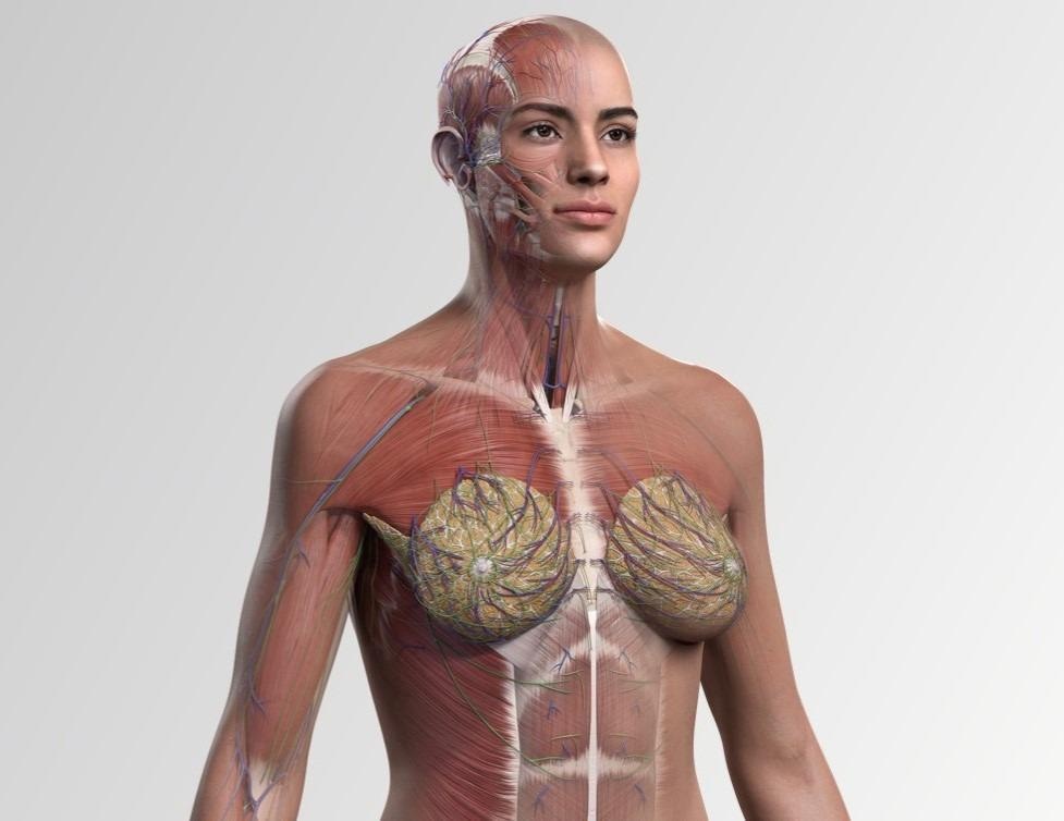
It might surprise you to know that the female anatomy has been historically underrepresented in the study of the human body since the 1500s. Our vision in developing this advanced 3D full female anatomy model is to create an experience where the male and female anatomy are equally represented. Our goal has always been to use the latest research, not just highlighting where the female differs from the male, as traditionally taught, but to promote equality through choice.
Ultimately, we want to provide the opportunity for educators around the world to teach an entire anatomy course using the female model as default, rather than the male, should they choose to. By providing this option, we want to play our part in disrupting the gender bias which exists in clinical practice.
Professor Claire Smith: Hello, I’m Professor Claire Smith, Head of Anatomy at Brighton and Sussex Medical School (BSMS). At BSMS we have been working with Elsevier for many years, and more recently we have been increasing the amount of diversity in our teaching. I was especially mindful of this when writing Gray’s Surface Anatomy and Ultrasound to ensure that we are teaching anatomy that is representative of all patients.
We are delighted that Complete Anatomy have developed the full female model and that we have been able to bring this into our teaching of medical students.
 The Complete Anatomy female model is the world's most advanced full female anatomy model. Image Credit: Elsevier
The Complete Anatomy female model is the most advanced full female anatomy model in the world and marks the first time that a female model has been built with such intricate detail in its entirety, versus replacing specific parts of male models with female anatomy. Why is this such a major milestone for education equality, and why do you believe this creation hasn’t been developed in the past?
Irene Walsh: One approach, which is regularly observed in the anatomy market, is to simply replicate the male skeleton and shrink it, adding in the female reproductive organs. Our team felt this would not align with our vision, it would not reflect the nuances of the female skeleton and how it presents, especially in anthropological instances. We wanted to represent the female as comprehensively as possible and set about researching points of sexual dimorphism seen in female bones using resources from forensic anthropology. We referenced anthropological data from specialists’ texts, academic papers, and customer feedback. This information was discussed in detail with subject matter experts. Once a brief was created on a specific region, a specialist medical writer worked closely with a 3D artist to craft the sexual dimorphic details apparent on the female.
Our team at Complete Anatomy from Elsevier is creating a platform of representation that is diverse, balanced, and most importantly, accurate. By giving educators the option to choose the female body as the basis for their students’ education, we’re helping evolve equitable teaching practices by normalizing the female body.
We are proud to take this step forward in addressing gender bias and believe our 3D full female model will have a clear impact on the educational experience of medical students worldwide as well as on the outcomes of patients they treat in the future.
Can you explain what the Complete Anatomy female model includes to create an accurate representation of female anatomy?
Irene Walsh: Informed by four years of expert research and development, anthropological data from specialist texts, academic papers, and customer feedback, our 3D full female anatomy model provides an unprecedented opportunity to study the female form in more detail than before.
The Complete Anatomy female model includes:
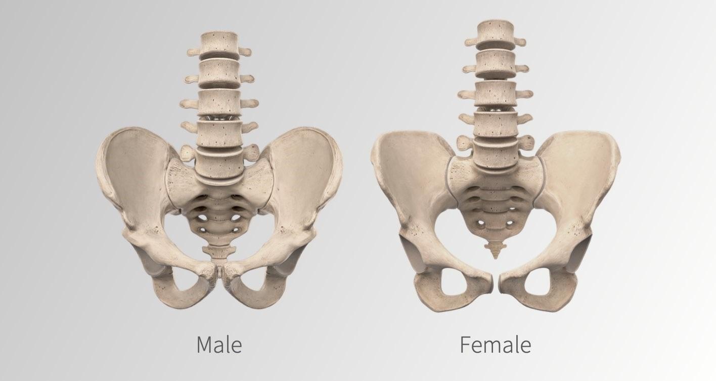
- The full skeletal system — A complete female skeletal system includes a wide array of unique features, rarely seen in anatomical texts. Sexual differences have been applied to areas such as the pelvis and skull. Long bones have been proportioned, and bone angles accurately reflect the uniquely female architectural skeletal base.
- Accurate portrayal of muscles — In line with research findings from the broadest demographic of females, each muscle has been refined, amounting to a reduction in the overall volume of muscle mass by roughly 30%.
- Visually detailed female-specific regions — The female-specific regions have been created in detail that is equivalent to the male counterpart. Breast tissue can be hemisected or quartered to reveal the underlying tissues with a more accurate distribution and representative state of the mammary glands, now shown as non-lactating, unlike many anatomical resources. The reproductive organs from the internal and external genitalia have been remodeled to accurately show their continued relationship.
- Comparative functionality — Users can switch between models for comparative study on any part of the male and female forms, compare sexual differences and reveal the origin and distribution of nerves. Users can learn from an interactive dissection course, take quizzes and watch videos to test their skills.
 Differences between the male and female forms are accurately portrayed via the Complete Anatomy female model. Image Credit: Elsevier
Why is it so important to have access to accurate models that can provide a complete understanding of the female anatomy?
Irene Walsh: With ‘Complete Anatomy’, we’re putting anatomy software in the hands of health science students on day one of their training. As a team, we take this responsibility very seriously. We are committed to playing our part in the changing and evolving nature of anatomical education in the 21st century, and our role in challenging previously held historical biases in the ongoing social conversations of our time.
We also believe it is important to have equal representation in the study of anatomy and remove unconscious bias that can be carried by learners into their future interactions with the body, including potentially with patients.
Professor Claire Smith: Traditionally, scientific understanding of anatomy and the generation of images in textbooks was based on the dissection of the human body. However, these dissections were predominantly performed on executed male prisoners. Therefore, these images and our understanding of the human body are based on the male anatomy, with elements of the female just added on as an adjunct. When half of the population is female, having just the breasts and pelvis added doesn’t reflect the comprehensive view of health care that is needed.
Accurate teaching that is removed from bias is really important from the start of medical education, as this enables the doctors and allied health care professionals of tomorrow to have an in-depth understanding of the human anatomy in both sexes.
 Complete Anatomy models allow students to utilize 3D technology alongside existing teaching methods for a deeper understanding of anatomical differences between sexes. Image Credit: valiantsin suprunovich/Shutterstock
Can you explain some of the practical applications of the model? Moreover, how is the Complete Anatomy female model currently being used to teach students?
Professor Claire Smith: At BSMS we use Complete Anatomy in a range of our teaching sessions. In lectures, the model enables 3D reconstruction of a specific area, in dissection sessions, this is available on iPads next to our amazing body donors so that students can compare the model to their donor. Additionally, in living anatomy and ultrasound sessions, students use the model to understand the difference between how the anatomy looks on themselves and then compared to without the skin on the model. Primarily the new female model helps us to pull out key points in each body to then analyze where there are differences and what the clinical significance of this is.
How does the model provide educators with a more comprehensive teaching approach? What are some of the benefits of using the female model in classes?
Professor Claire Smith: For the first time this model has provided us with a full resource of the female form, rather than just a male skeleton with a female pelvis. The benefits of this range beyond just the overt; such as the in-depth exploration into the anatomy of the breast; it has also given us resources to explore many more subtle elements of the female anatomy such as the difference in long bones. The model has also contributed to the hidden curriculum as it demonstrates equality in our teaching and creates awareness of the historic bias in medicine.
Introducing the full Female model: Complete Anatomy 2022
The Complete Anatomy female model signifies a major milestone in equal representation, however, there is still a long way to go. How do you believe we can further improve education equality in terms of the underrepresentation of the female anatomy in the study of the human body?
Irene Walsh: Complete Anatomy is just one small segment of a massive project being undertaken across the teams at Elsevier, a global leader in research publishing and information analytics, with the potential to significantly impact learning and teaching globally. We are passionately committed to increasing inclusion, diversity and equity in research, healthcare, and publishing, and in particular to increasing the representation of under-represented groups, including women, people of color, and socially disadvantaged populations, among our editorial advisors, reviewers, authors, and contributors. We are working together to promote a stronger, more diverse, and representative scholarly community of authors, editors, and contributors built on a platform of inclusion and diversity.
Professor Claire Smith: I believe that we need to work alongside the national curriculum to develop more of an open and inclusive approach when delivering teaching on anatomy. By working to remove the historical bias of anatomy education, students of both sexes can gain a more comprehensive understanding of female health. This also extends beyond the underrepresentation of female anatomy to include better education on differences in age, race, normal variation, and body type.
Where can readers find more information?
Irene Walsh: www.3d4medical.com/
Professor Claire Smith: https://www.bsms.ac.uk/about/contact-us/staff/professor-claire-smith.aspx
About the Interviewees
 Irene Walsh: In her role at 3D4Medical from Elsevier, Irene Walsh leads the Product, UX, Medical Content, and 3D teams to deliver engaging medical teaching and learning experiences, through the use of 3D Anatomy.
In 2015, Irene became only the ninth woman to present on the Apple Keynote stage, demoing the company’s flagship product Complete Anatomy to an estimated audience of over 40 million. Her passion lies in user-centered product design and tackling the biases in anatomical learning, through developing great experiences and leveraging cutting-edge technology. Irene has lectured at Technical University Dublin in Design Practice, held workshops on Tackling Bias in Education, and is a council member of the National Design Forum of Ireland.
Professor Claire Smith: As a Professor and Head of Anatomy, I oversee all aspects of teaching to over 1,500 medical students and allied health professionals in a year. This includes leading a body donation program of which at BSMS alone we require 60 donors a year. My career highlights include being awarded National Teaching Fellowship, and on a daily basis having great conversations with students and surgeons really motivates me to deliver excellent teaching programmes.
For more information on body donation please visit: https://www.hta.gov.uk/guidance-public/body-donation/how-donate-your-body