Can surgeons quantify the risk of aphasia when removing a brain tumor? To find out, researchers at Klinikum rechts der Isar of the Technical University of Munich (TUM) are analyzing the brain as a network. In a current study with 60 patients, they already achieved an accuracy rate with three quarters of their predictions.
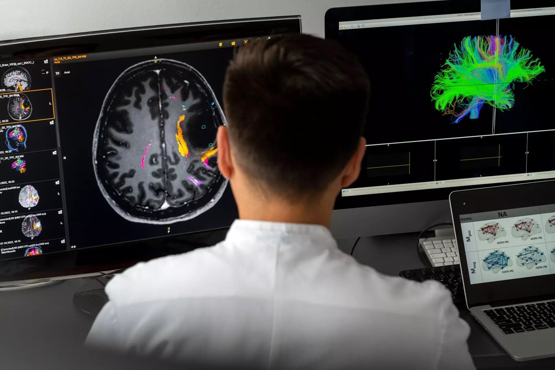
 With a special form of magnetic resonance tomography known as tractography scientists of TUM produce 3D representations of the networks of neural pathways in the brain. Image Credit: Technical University of Munich (TUM)
Brain tumors are relatively rare. According to the German Society for Neurology, the annual incidence is approximately five cases per 100,000 inhabitants. “But in most cases the surgical removal of the tumor is unavoidable,” says Prof. Sandro Krieg, who estimates that a glioma – a common type of brain tumor – is removed at Klinikum rechts der Isar of the Technical University of Munich (TUM) “on an almost daily basis”.
Depending on the tumor, Krieg and his colleagues develop individual treatment and surgical strategies. A crucial point: healthy tissue should be preserved to the greatest possible extent and no structures should be damaged that could lead to further limitations. “Aphasia” is the term for post-surgical speech impairments, for example. “We want to have precise knowledge of the risk of aphasia prior to the operation.”
The chief physician at the Neurosurgery Clinic at Klinikum rechts der Isar has been studying preoperative brain mapping for more than 10 years. “For a long time we have known the basic locations in the brain responsible for such functions as movement or speech. But it is only in the past five years or so that we have begun to analyze the brain network to learn how the various regions work together, for example to enable a person to speak. One thing is clear: there is no language center as such. Instead, the structure is more like several hubs or nodes of a big network through which speech is made possible.”
Brain tumor: making predictions through machine learning
Analyzing the network characteristics of the brain – referred to as connectome analysis – a process used by Prof. Krieg’s team for around two years – plays a key role in current research. “In this way we quantify the connections in individual brain regions,” says Prof. Krieg. “We have since started to assign more precise functions to brain regions.”
TUM scientists Dr. Haosu Zhang and Dr. Sebastian Ille have anatomically mapped images of brain layers responsible for speech capabilities. The process is as follows: “With a special form of magnetic resonance tomography known as tractography, we produce 3D representations of the networks and subnetworks of neural pathways in the brain,” explains Zhang.
This network analysis is supported by the process of navigated transcranial magnetic stimulation, in which a targeted magnetic pulse inhibits nerve cells in fiber pathways responsible for speech. This causes a temporary speech impairment in the patient that can be recognized in video analysis. It enables researchers to precisely identify brain regions responsible for speech.
We combine the so-called connectome parameters from tractography with information on the patient’s speech function."
Dr. Haosu Zhang, TUM Scientist
What makes Zhang’s and Ille’s algorithm special: it yields “statistically significant parameters” – data that can be used to train a machine learning model and thus to localize the speech of individual patients. As complex as the use of the different analysis methods may seem – the defining feature of the method is its simplicity: the entire analytical process works without complex algorithms or powerful computers. “The data that we use are obtained from routine hospital tests,” says Zhang.
Network analysis: 73 percent accuracy in predicting speech impairments
In a recent study with 60 patients, the researchers at Klinikum rechts der Isar showed that this combined analysis can predict with considerable accuracy (73%) whether surgery will cause speech difficulties (post-operative aphasia). “It is very important to be able to make these predictions,” says Krieg. He is excited by the possibility of being able to quantify the risk more precisely by means of “real network analysis” and having concrete data to support the mapping of the brain.
Moreover: with the help of machine learning, the predictions will become even better over time. But for that purpose the researchers will need more patient data in order to train the machine learning algorithms. “It is the only approach that can use big data to predict the risk of a surgical intervention,” says Prof. Krieg, who now plans to find more patients to participate in his research. He believes that even “a few hundred” patients will be enough for highly precise predictions.
Source:
Journal reference:
Ille, S., et al. (2022) Preoperative function‐specific connectome analysis predicts surgery‐related aphasia after glioma resection. Human Brain Mapping. doi.org/10.1002/hbm.26014.