In this interview conducted at Pittcon 2023 in Philadelphia, Pennsylvania, we spoke to Ron Heeren, a speaker at the 2023 James L. Waters Symposium.
Please could you introduce yourself, and tell us about your personal background and what first attracted you to this field?
I am Ron Heeren, a distinguished professor of molecular imaging at Maastricht University in the Netherlands. I am also the director of the Maastricht MultiModal Molecular Imaging Institute. I was trained as a physicist, then developed a career in biochemistry, and now I am teaching in a medical center.
I have always wondered about the complexity of nature. One of my heroes, Richard Feynman, once said that you have to stop and think about it to really appreciate nature’s complexity; the inconceivable nature of nature.
I love that quote because it describes what triggered me to go into science: to satisfy my curiosity and understand the complexity of the world around me. The beauty of molecular imaging is that it does precisely that. It shows the inconceivable complexity of nature on a microscope slide.
Unlocking Nature's Secrets with Advanced Molecular Imaging Techniques
What is secondary ion mass spectrometry?
The field I am engaging in is molecular imaging using mass spectrometry. There are essentially two ways of generating images with a mass spectrometer. The first is firing lasers at a surface to evaporate and ionize molecules, then analyzing them in the mass spectrometer. The second uses an ion beam, where a primary ion generates secondary ions that are then analyzed by a mass spectrometer.
The latter is the field of secondary ion mass spectrometry. In my work, we use both in concert because each technology has complementary features. The beauty of SIMS is that it can achieve spatial resolutions, such as no other technique in imaging mass spectrometry.
What is molecular imaging more broadly and what are its advantages?
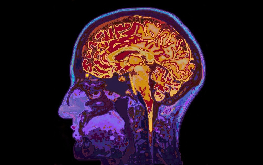
Molecular imaging is a form of molecular photography where we take snapshots of the molecules on highly complex surfaces, such as tissue sections or biopsies of cancer patients, solar cells, or even leaves with microbes growing on them, and we try to visualize them.

Image Credit: SpeedKingz/Shutterstock.com
Molecular imaging produces a map or a photograph of the spatial location of the molecules combined with the identity of the molecules themselves.
How can secondary ion mass spectrometry be employed in molecular imaging?
The beauty of secondary ion mass spectrometry is its incredible spatial resolution. These ion beams can be focused down to an extremely small spot, down to 50 nanometers. With molecular imaging, we can generate very small pixels, which provides an insight into what is going on in a single cell in the context of a complete tissue.
Essentially, SIMS brings very high spatial resolution. One added advantage is that we can study an individual single cell, layer by layer, and create a three-dimensional map of all molecules in that single cell.
In what medical fields can advances in digital molecular pathology have an impact?
Molecular imaging is entering digital pathology, a pathologist looking at digital images rather than through a microscope. As molecular imaging using mass spectrometers generates digital images, they can be shared easily with the pathologist.
They can be layered on top of the optical images that they already have. Now, the pathologist can augment how they look at the problem with molecular information. One example is detecting tumor cells in biopsy tissue. We can look at tissue sections from cartilage from damaged knees to understand the healing process and design new drugs.
We can look at pharmaceutical and animal models, where we observe where the drug ends up, how it is metabolized, and if it has an effect. These are all problems in a spatial context. These technologies can be applied with a gold star in biomedicine and pharmaceutical research.
How can innovative imaging technologies offer new insights into life's complexity?
We see these images in more molecular detail as our mass spectrometers improve. Some of these molecular changes that trigger a disease process or that people want to interfere with when designing a new drug (to circumvent a disease) are related to minute molecular changes.
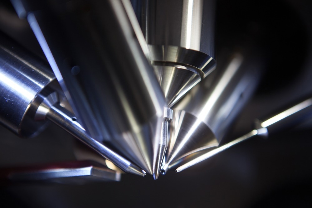
Modern mass spectrometers enable us to see things like isomeric species. This lipid has a double bond very close to the glycerol backbone or very far away, two very different structures. We can now visualize where that structure differs in a tissue or cell.

Image Credit: Intothelight Photography/Shutterstock.com
Mass spectrometry imaging can unravel this complexity at spatial and molecular detail levels. We can look at the identity of a molecule, where the molecule is in the cell, where that cell is in a piece of tissue, and where that tissue comes from in a patient. This enables the description of the entire translational imaging chain.
The 34th James L. Waters Symposium highlights the development, commercial construction, and recent advances in instrumentation and its applications. What are some of the recent advancements in secondary ion mass spectrometry?
The James L. Waters Symposium highlights instrumentation advances, such as targeted pathology, where people use labeled antibodies to observe targeted proteome processes in detail. Another technology presented that we worked on is using a detector from CERN in the molecular pathology field to accelerate the rate at which we can generate these images.
We would take perhaps a hundred to a thousand pixels per second on a typical commercial instrument. Each pixel corresponds to the mass spectrum. These new detectors from CERN enable the acquisition of a millions of pixels per second, so we are very close to achieving our current goal of scanning one tissue slide in one minute.
This will be perfectly in sync with the pathology workflow. Our molecular imaging technologies with SIMS and CERN would seamlessly fit into the digital pathology workflow.
How important is it to understand the history of the important contributions and cooperations in this field?
The importance of history cannot be stressed enough because we all stand on the shoulders of giants. One of the approaches that we are still working with was developed in the 1960s, but now we have leapfrogged away with all the new technologies that were not available back then, but the basic ideas remain the same.
Unraveling Nature's Complexity with Molecular Imaging - Ron Heeren at Pittcon 2023
These early ideas are now accelerating because of all the new available technologies. This symposium beautifully highlighted that as it brought together all these different elements: instrumentation, engineering, application in the clinic, targeted pathology, and the history on which it all was based.
What are the current challenges within the secondary ion mass spectrometry-based molecular imaging field?
One of our biggest challenges is the sheer amount of data we produce. One aspect of this is data storage, as we are obliged by law to keep patient data for up to 15 years. If I generate three terabytes in 10 minutes and store it for 10 or 15 years, then my storage bill will outweigh my electricity bill very quickly.
Researchers are looking into smarter solutions for storing data, maybe only acquiring relevant data to reduce the amount of data we generate.

Image Credit: Gorodenkoff/Shutterstock.com
The other aspect is what we do with the data and how we interpret it. We have a million spectra per second; no human mind could go through these spectra individually. We need more innovative artificial intelligence, machine learning, and neural network tools to explore the data and find the relevant information we seek to understand the complexity of health and disease.
How do you hope your work will help overcome some of the challenges you mentioned?
I am afraid my work will only worsen those two challenges because we are generating more data in a shorter time. However, while we are doing that, we also face these challenges. We have many bioinformaticians that we collaborate with to tackle those problems using machine learning and neural networks.
I think my team’s contribution to this field is that we see that to solve these challenges, we need researchers from many different disciplines. It is not just the tool we develop to solve data analysis challenges but how to collaborate across the boundaries of disciplines.
This is one thing that imaging mass spectrometry excels at because there is the fundamental side, instrumentation, application development, and data handling. They all have to come together. Our contribution is bringing these people together in the institute around the appropriate infrastructure to tackle these challenges.
What are you currently working on that you are particularly excited about?
One project we are excited about is single-cell imaging using our CERN-based detector to build libraries of molecular profiles of immune cells and then automatically recognize these cells in a piece of tissue.
This allows us to understand how the metabolic phenotype of an immune cell changes in the presence of a tumor and as a response to the distance to the tumor. So much more complexity is yet to be discovered and understood, which will significantly contribute to that.
About Professor Ron Heeran
Prof. Dr. Ron M.A. Heeren obtained a PhD degree in technical physics in 1992 at the University of Amsterdam on plasma-surface interactions.  He started to work on molecular imaging instrumentation and its application as a research group leader at FOM-AMOLF, Amsterdam. In 2001, he became professor at the chemistry faculty of Utrecht University lecturing on the physical aspects of biomolecular mass spectrometry. In 2014 he started as distinguished professor and Limburg Chair at Maastricht University. He is the founder and scientific director of M4I, the Maastricht MultiModal Molecular Imaging institute on the Brightlands Maastricht Health campus. He was awarded the prestigious 2019 Physics Valorization prize by the Dutch organization for scientific Research, NWO and the 2020 Thomson medal of the international mass spectrometry foundation. In 2021 he was elected as a member of the Royal Dutch Academy of Sciences, KNAW. His academic research interests are mass spectrometry based personalized medicine, translational molecular imaging and “omics” research, high-throughput bioinformatics and the development and validation of innovative molecular analytical imaging techniques across the scientific disciplines.
 About Pittcon
Pittcon is the world’s largest annual premier conference and exposition on laboratory science. Pittcon attracts more than 16,000 attendees from industry, academia and government from over 90 countries worldwide.
Their mission is to sponsor and sustain educational and charitable activities for the advancement and benefit of scientific endeavor.
Pittcon’s target audience is not just “analytical chemists,” but all laboratory scientists — anyone who identifies, quantifies, analyzes or tests the chemical or biological properties of compounds or molecules, or who manages these laboratory scientists.
Having grown beyond its roots in analytical chemistry and spectroscopy, Pittcon has evolved into an event that now also serves a diverse constituency encompassing life sciences, pharmaceutical discovery and QA, food safety, environmental, bioterrorism and other emerging markets.