In neonates and children, cardiovascular and gastrointestinal problems are the significant causes of death in the first five years of life. Using continuous monitoring systems helps guide clinical decisions. Current hospital systems use sensors, cables, and wires connected to monitors. However, fortunately, advances in bioengineering have led to the development of wireless, skin-interfaced sensors for use in the simultaneous acquisition of different classes of signals.
Digital stethoscopes, including wearable designs, can provide (complementary) information on airway obstruction, intestinal motility, cardiac activity, and adventitious lung sounds. However, they cannot be used for continuous monitoring due to some limitations, and consequently, the clinical use of body sound typically occurs through periodic measurements.

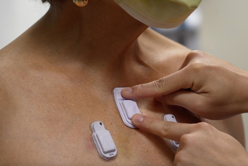
A health care worker places the wearable devices across a patient's chest to capture sounds throughout the lungs that are associated with breathing. Image Credit: Northwestern University, Study: Wireless broadband acousto-mechanical sensing system for continuous physiological monitoring
The study and findings
The present study introduced wireless BAMS systems for continuous physiological monitoring. The BAMS system can capture a broad range of signals, from slow body movements (around 0.01 hertz [Hz]) to high-frequency body sounds (up to 1 kHz). Gentle adherence of the device at the suprasternal notch could allow for simultaneous measurement of respiratory and cardiac sounds.
Placing time-synchronized devices on the abdomen could enable spatiotemporal monitoring of gastrointestinal sounds. In an advanced implementation, 13 devices could be placed across target sites across the posterior and anterior chest to monitor pulmonary health, disease progression, and rehabilitation. This (advanced) implementation can be applied to patients of any age, including newborns admitted to the neonatal intensive care unit (NICU).
The BAMS device includes body- and ambient-facing microphones, an inertial measurement unit, a flash memory, a wireless-charging antenna, and a standard Bluetooth low-energy system-on-a-chip mounted on a printed circuit board. The microphones capture sound from two directions, and an adaptive filtering algorithm minimizes the contribution of body sounds to ambient sounds and vice versa.
The BAMS system can be used in daily life scenarios, allowing for the monitoring of standard parameters (respiratory rate, heart rate) and autonomic measures, such as cardiorespiratory coupling, swallowing, and heart rate variability (HRV). The system can operate across various activities, such as sleep and exercise. Besides, the researchers compared the BAMS data of a neonate admitted to NICU to readings from Food and Drug Administration (FDA)-approved clinical monitors.
Sound intensity and breathing interval determined by the BAMS device correlated with pauses in breathing and airflow rate. In addition, the device demonstrated reliable monitoring of heart rates, respiratory sounds, and other parameters over a protracted period (of three hours) in a cohort of five neonates admitted to the NICU. Respiratory sounds aligned well with chest movements and data from respiratory inductance plethysmography and nasal temperature.
Further analyses showed that bowel sounds captured by BAMS devices were correlated with electromyography signals from an adult’s abdomen. Additionally, the researchers used 13 devices mounted on 35 chronic lung disease patients and 20 healthy individuals. Data from a healthy subject showed similar distributions of sound intensities, chest wall movement, and sound frequencies from the right and left body sides.
Similar measurements on patients with surgical lung resections and those with chronic lung diseases reflected their condition. One participant with resection surgery of the left upper lobe and right lower and upper lobes showed lower pulmonary function in the removed lobes, reducing the sound intensity and airflow rates.
A comparative analysis of data from healthy subjects and chronic lung disease patients highlighted the importance of airflow volume, sound frequency, and airflow rate in diagnosing restrictive and obstructive lung diseases. These results relied on data from BAMS devices placed on the (lower and upper) posterior regions of the chest and suprasternal notch, with separate airflow rate and flow volume measurements using a peak flow meter.
Sound energy could be additionally calculated by integrating sound intensity over time. These parameters could help with tracking disease progression and treatment response. Air volume and airflow rate measurement can also facilitate monitoring the Tiffeneau-Pinelli index. Sound intensities at the suprasternal notch were higher in healthy participants, with an average intensity of 54 decibels (dB) than in chronic lung disease patients.
However, the average intensities were 38 dB in chronic lung disease patients without lung resection, 36 dB in patients with right upper lung resections, and 30 dB in subjects with left upper lung resections. The right upper posterior dominant expiratory frequency was 219 Hz in healthy participants and 256 Hz in chronic lung disease patients. Further analyses at different locations of the lungs revealed stark differences between chronic lung disease patients and healthy subjects.
Conclusions
The study presented technology for simultaneous measurements of body sounds and movements as a source of physiological signals, with home and hospital applicability. Numerous characterization studies and benchmarking measurements confirmed the accuracy of the BAMS system. Overall, the combination of a microphone pair, sound-separation algorithm, time-synchronized operations, and small skin-interfaced form creates unique possibilities for (continuous) patient monitoring.