Microplastics are synthetic polymer particles ranging in size from one micrometer (μm) to five millimeters (mm). They are abundant in all environments, including air, water, soil, and the food chain.
Recently, microplastics have been detected in various human tissues and organs, including lung, colon, liver, placenta, breast milk, testes, blood, urine, and stool. Emerging evidence indicates that high-level exposure to microplastics may lead to inflammation and oxidative stress, both of which are major hallmarks of many non-communicable chronic diseases, including inflammatory bowel disease (IBD).
Endometriosis is a chronic and inflammatory gynecological disease characterized by the presence of endometrial-like tissues outside the uterus. Although the exact etiology of endometriosis remains unclear, it is generally believed that a complex crosstalk between genetic, environmental, hormonal, and immunological factors is associated with the development of this condition.
Study findings
In the current study, scientists assess the presence of microplastics in urine samples collected from healthy individuals and those with endometriosis. To this end, the researchers characterized the identified microplastic particles using micro-Fourier Transform Interferometer (μFTIR) spectroscopy and scanning electron microscopy with energy dispersive X-ray spectroscopy (SEM-EDX).
A total of 38 human urine samples, which included 19 healthy donor and 19 endometriosis patient samples, were analyzed, along with 15 pre-filtered water samples, which served as procedural blanks.
Characterization of microplastic particles in urine
The analysis of healthy donor urine samples led to the identification of 23 microplastic particles consisting of 22 polymer types in 17 samples. Comparatively, the analysis of endometriosis patient urine samples led to the identification of 232 microplastic particles composed of 16 polymer types in 12 samples.
The average levels of microplastic particles in healthy donor and endometriosis patient urine samples were 2,575 and 4,710 particles/liter, respectively. The most abundant polymer types present in healthy donor samples were polyethylene (PE), polystyrene (PS), resin, and polypropylene. In endometriosis patient samples, polytetrafluoroethylene (PTFE) and PE were the most abundant polymer types.
The mean length and width of microplastic particles detected in healthy donor samples were 61.92 and 34.85 μm, respectively. About 66% and 30% of particles were fragments and films of clear or white color, respectively.
The mean length and width of microplastic particles detected in endometriosis patient samples were 119.01 and 79.09 μm, respectively. About 95% of particles were fragments, 4% were films, and less than 1% were fibers. About 96% of the particles were clear or white.
The metal catheter-derived microplastic particles from endometriosis patients and healthy donor-derived microplastic particles were significantly smaller than those detected in procedural blanks.
A total of 62 particles consisting of 10 polymer types were detected in procedural blanks. The most abundant polymer types were PP, PE, and PS. Thus, the presence of contaminants cannot be avoided while measuring microplastic particles in donors.
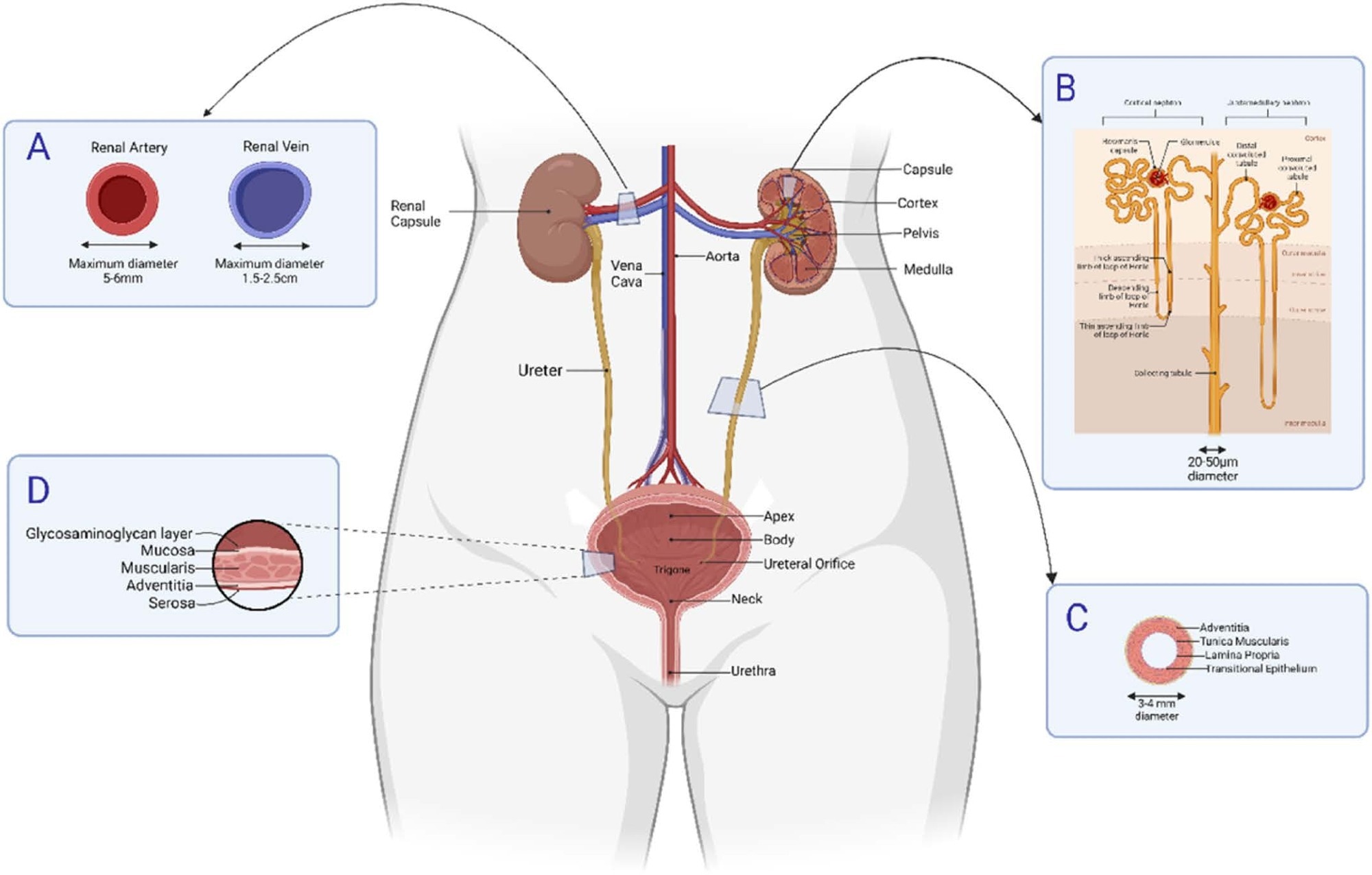
 Proposed MP transport to the bladder relative to the diameters of the internal tubules and associated blood vessels. A) renal arteries, branching from the aorta, and the renal veins drain into the inferior vena cava. The kidney is a fibrous capsule that can be divided into renal parenchyma consisting of two layers, a renal cortex and the renal medulla. The renal medulla consists of 10–14 renal pyramids (B), each separated renal columns where urine is made. Urine drains through the minor/major calyx and into the ureter (C). The ureter transports urine to the bladder. Its blood supply is segmental and includes the renal, common iliac and internal iliac arteries. Urine collects in the urinary bladder (D), which can hold 400–600 mL urine. During urination, the bladder contracts allowing urine to be expelled via the urethra. The bladder’s main blood supply is the superior vesical branch of the internal iliac arteries with venous drainage into the internal iliac veins. (Created in BioRender.com).
Characterization of non-microplastic chemicals
Several non-microplastic chemicals, such as microplastic building block polymer monomers or polymer additives, were detected in urine samples derived from healthy donors and endometriosis patients.
N-(2-ethoxyphenyl)-N-(2-ethylphenyl)-ethanediamide was the most abundantly detected chemical, followed by bisphenol A propoxylate/ethoxylate, bis(2-hydroxyethyl) dimerate, and tetra-ammonium octa-molybdate. The most abundantly detected non-microplastic particles were cellulose fragments and zein fragments.
Conclusions
Microplastic particles were identified in urine samples collected from both healthy individuals and endometriosis patients, with no significant differences in the levels of microplastics observed between the two groups.
A high level of PTFE fragments has been detected in urine samples from endometriosis patients. PTFE, also known as Teflon, is widely used as a non-stick coating and lubricant in cookware, car interiors, and dental floss.
Teflon used in surgical applications has been found to cause Teflon granuloma, which is an inflammatory giant-cell foreign body reaction to PTFE fiber exposure. Additionally, workplace exposure to PTFE particles has been associated with small airway-centered granulomatous lesions.
PTFE belongs to the polyfluoroalkyl substances (PFAS) group of chemical contaminants known to cause immune suppression, thyroid dysfunction, liver disease, lipid dysregulation, and endocrine disruption in humans. High-level exposure to PFAS through drinking water has been associated with an increased risk of ovary syndrome, uterine leiomyoma, and infertility.
The larger and irregularly shaped microplastic particles identified in urine samples from endometriosis patients highlight the possibility of developing inflammatory and oxidative stress-related responses, which might impact the development of endometriosis.
The size and shape of microplastic particles detected in urine raise uncertainty about their transportation through the small capillary networks of the kidneys to reach the bladder. Further experiments are needed to determine the route of uptake and transport of microplastic particles throughout the human body and the health consequences of microplastic exposure.
Journal reference:
- Rotchell, J. M., Austin, C., Chapman, E., et al. (2024). Microplastics in human urine: Characterisation using μFTIR and sampling challenges using healthy donors and endometriosis participants. Ecotoxicology and Environmental Safety. doi:10.1016/j.ecoenv.2024.116208