Introduction
The Québec Consortium for Drug Discovery (CQDM), OSÉO and Alsace-Biovalley have jointly funded a project called the Imaging and biosimulation platform for new central nervous system drugs (BICNSD). This international collaboration between France and Québec is aimed at achieving the following:
- To develop and verify synergistic platforms which can be used to render an exclusive service based on imaging and biosimulation
- To advance discovery and development of drugs via improved understanding of diseases relating to the central nervous system (CNS)
- To track the effects of drug candidates at both molecular and subcellular levels
Under this 3-year initiative, efforts will be made to develop innovative processes and technologies that will advance the discovery and development of novel drugs. The two synergistic platforms include the multiplex cellular fluorescence imaging platform and the biosimulation platform for CNS drug delivery. The former will be handled by Photon etc., while the latter will be managed by Rhenovia Pharma.
For this project, Photon etc. will partner with Professor Paul de Koninck who serves at the Neurophotonics Centre of Université Laval based in Québec.
Project Objectives
The basic mechanisms of CNS diseases are still not defined properly, mainly due to insufficient methods for studying complex processes at the synapse level and the poor outcomes of the available treatments. In order to overcome these issues, advanced tools are required to study protein dynamics along the neuronal membrane, subsequent to different drug treatments or stimulations.
The international project aims to discover and develop a platform for CNS diseases based on several parameters, including:
- A cellular and subcellular imaging solution that can simultaneously image a number of labels
- A probe design for labeling receptors in neurons
- A biosimulation platform that implements and combines signaling cascades, specific interactions between proteins, and movement of receptors along the postsynaptic membrane
- Software tools for Image analysis and visualization of labels
Biosimulation Platform
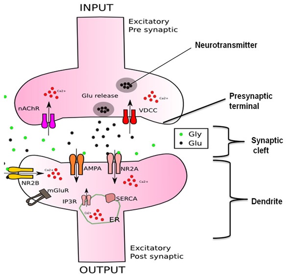
Synapses in the nervous system (Figure 1) allow neurons to transmit chemical or electrical signals to one another. Synapses comprise three elements: the presynaptic terminal, the synaptic cleft, and the postsynaptic element.

Figure 1. Structure of the synapse
The neurotransmitter released by the presynaptic terminal triggers receptors, which in turn convert the chemical signal into biochemical and electrical signals by triggering intracellular secondary messenger pathways and ion channels.
Neurotransmitter receptors have specific properties such as affinity for their kinetic and ligand characteristics like deactivation, activation, and desensitization that considerably impact their activation profile.
Besides these intrinsic specificities, the location within the synapse that is close (synaptic) or far (extra synaptic) from the release site will also impact the response.
The initial step in the study was to investigate the dynamics of proteins and the interactions of glutamate receptors such as NMDA, AMPA and metaboprobic glutamate receptor (mGluR) as well as their subtype of receptors such as mGluR5, GluA1 and GluA2 AMPAR, NR1/NR2B NMDAR, and NR1/NR2A, along with intracellular proteins of interest with respect to the signaling pathway.
A literature review was later performed to establish whether these receptors relocated due to the effect of drug treatment or due to specific pathological conditions. To this end, models of NR1/NR2B receptors were developed, verified, and then applied in the glutamatergic synapse with specific characteristics.
Goals for the future will include the characterization of the remaining glutamate receptors. For this purpose, Photon etc. has developed unique cameras to observe these characteristics and determine the computational environment. In addition, dendritic branches will be studied and integrated with around 10-15 excitatory synapses to get a clearer picture.
Multi-labeling of Receptors in Neurons
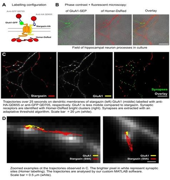
Using different combinations of antibodies and quantum dots, multi-labeling of two types of synaptic receptors (NMDA and AMPA) was carried out (Figure 2). Also, labeling of post-synaptic sites on living neurons was performed.

Figure 2. Specific labeling of two types of synaptic receptors AMPA and NMDA.
The laboratory is also implementing a new method to measure the correlation and co-localization of proteins that are labeled differently so that they serve as a proxy for interactions between proteins.
Multiplex Cellular Imaging Platform
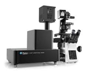
In an attempt to overcome the limitations of conventional labels used for studying and imaging different molecular species and tissues, Photon etc. developed a unique technology called a spectral imager (Figure 3) that uses innovative narrow band labels like SERS nanospheres and quantum dots and enables multiplexing of various labels, thereby monitoring and imaging various signals at the same time. This way, detailed in vitro studies can be carried out when exploring the effect of novel drug candidates on signalization cascades at the cellular level.

Figure 3. The system is anticipated to allow the acquisition of spectrally resolved fluorescence images within a few seconds
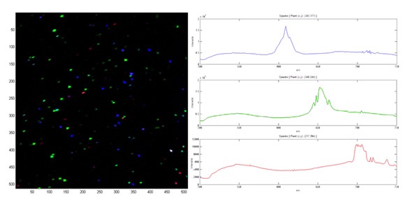
The prototype design for fluorescence cellular imaging has now been accomplished, and the production model is presently being developed. The system includes Photon etc.’s hyperspectral imager integrated with Nüvü Cameras’ HNü 512 EMCCD camera and an IX-73 Olympus microscope. Figure 4 shows the initial characterization of quantum dots made at 605, 655 and 705nm wavelengths.

Figure 4. Spectra of quantum dots at 605nm (blue), 655nm (green) and 705nm (red)
The filter is specifically designed for rapid acquisition of hyperspectral data at 500 to 900nm wavelength range with a bandwidth of 2nm. Optical filters from Semrock and a 300W xenon lamp from Sutter Instruments provide the required illumination. Equipped with mirror turret and motorized focus, the microscope allows for automatic selection of illumination wavelength and the corresponding z-stack acquisitions.
Conclusion
The BICNSD project will not only help in advancing the discovery and development of new drugs for CNS diseases, but will also improve our understanding of these diseases at both molecular and subcellular levels.
About Photon etc
 Photon etc. offers state-of-the-art photonic and optical research instrumentation, from laser line tunable filters to widefield and microscopy hyperspectral imaging systems. Its patented spectral imaging and optical sensing technologies provide solutions for a wide variety of scientific and industrial applications. From material analysis to medical imaging, Photon etc.’s expertise and spirit of innovation allow the exploration of uncharted territories.
Photon etc. aims to provide each researcher, engineer and technician with access to the latest innovations in optical and photonic instrumentation. As pioneers in Bragg-based hyperspectral imaging, Photon etc. offers state-of-the-art instruments, driven by its clients’ desires to surpass limitations in measurement and analysis.
Inspired by the scientific creativity found in Montreal and Quebec, Photon etc. promotes open and collaborative innovation and excellence. The dynamic team of this company is proud to offer to its clients innovative and reliable instruments, based on the latest scientific advances in photonics and optics. At Photon etc., the primary wish is to develop a long term relationship with the clients by providing products adapted to their specific needs, combined with personalized service and support.
Sponsored Content Policy: News-Medical.net publishes articles and related content that may be derived from sources where we have existing commercial relationships, provided such content adds value to the core editorial ethos of News-Medical.Net which is to educate and inform site visitors interested in medical research, science, medical devices and treatments.