Sponsored Content by Metrohm AGReviewed by Olivia FrostMay 20 2024
The medical field is a new frontier for Raman spectroscopy. Raman has already been employed in dentistry and cancer investigations, and it is now expanding into Point-of-Care (POC) applications.
This article provides a summary of some novel and intriguing studies into the use of Raman spectroscopy for the identification of malignant tissue, disease biomarkers, and disease-causing pathogens.
Detecting bone infection with a handheld Raman spectrometer
Infection complications are a big risk when using human bone grafts in musculoskeletal surgery. Bone-related infections are typically caused by Staphylococcus epidermidis and Staphylococcus aureus, which can be challenging to treat.
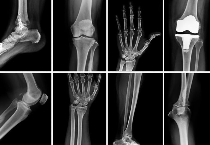
Image Credit: Adobe Stock
Detecting staph bacteria on graft material, as well as distinguishing between healthy and infected bone, are critical for infection prevention. Lab culture findings typically take 7–10 days and are susceptible to contamination during transport and testing.
In positive cases, the patient must be treated retroactively with high antibiotic dosages. On-site analysis allows the surgical team to identify and prevent diseased bone on the spot, making it an appropriate solution to this problem.
A study group in Austria has recently demonstrated a successful distinction between healthy and diseased bone samples, as well as between two strains of staph bacteria, using a portable MIRA Raman spectrometer.1
Their method distinguished between healthy and diseased bone by analyzing the fingerprint Raman bands of phosphates, amides, and collagens and their varying intensity and peak width ratios. Principal component analysis (PCA) aided optical analysis and was used to distinguish between staph strains more precisely.
The Austrian group admired MIRA’s compact, lightweight, and battery-powered design, along with the fact that Raman spectroscopy requires minimal sample preparation and produces fast results.
Testing required only a tiny bone sample for in-situ examination during surgery, and fast and accurate results were provided directly in the operating room.
Raman spectroscopy for cancer detection
Raman’s molecular fingerprint spectra are sensitive enough to identify disease-related chemical changes. Surgeons can use compact Raman spectrometers to examine tumors during surgical procedures, allowing for quick decision-making. The sections below include application examples for breast and pancreatic cancer.

Image Credit: Adobe Stock
Traditional Raman spectroscopy in breast cancer assessment
When breast cancer is suspected, patients often undergo one operation for a biopsy and a subsequent one to remove malignant tumors if confirmed. However, the ability to assess suspect tissues during the initial surgery could allow for the immediate removal of any necessary tissues.
The benefit to both the patient and the medical sector is immeasurable, and research indicates that Raman spectroscopy may be able to satisfy this demand for some forms of cancer.
Raman spectroscopy is sensitive enough to identify tissue changes caused by several forms of cancer. For instance, there are very subtle differences in Raman spectra from healthy breast tissue and malignant tumors.
Researchers in the United Kingdom employed a high-resolution i-Raman laboratory apparatus (Figure 1) and multivariate methods, including PCA, to successfully discriminate between healthy and malignant tissues.2
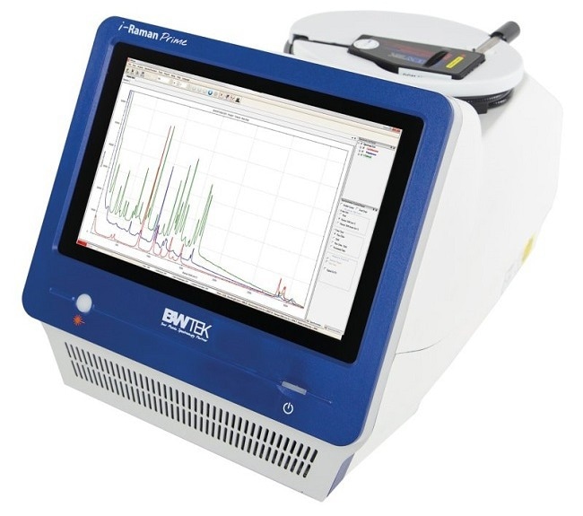
Figure 1. The i-Raman Prime 785S Portable Raman Spectrometer from Metrohm. Image Credit: Metrohm AG
SERS for detection and measurement of pancreatic cancer biomarkers
Surface-enhanced Raman spectroscopy (SERS) can serve as an effective alternative when standard Raman spectroscopy is unsuitable for analysis. This is particularly relevant in cases where the target molecules are present in a complex sample matrix or where the fluorescence of carbon-based molecules interferes with the analysis.
SERS amplifies the Raman signal without enhancing the competing fluorescence, making it more effective. Additionally, SERS enables the sensitive detection of analytes at concentrations as low as mg/L and, in some instances, down to µg/L. The peaks in SERS are sharp and well-defined, facilitating the efficient detection and identification of target analytes.
Pancreatic cancer is fatal, in part because it is difficult to diagnose. However, some biomarkers exist at elevated levels in ~75 % of positive cases.3 Enzyme-linked immunosorbent assays (ELISA) can identify these biomarkers, which include antibodies, antigens, and proteins.
In an emerging approach, an i-Raman laboratory spectrometer at the University of Utah was used for SERS analysis in combination with ELISA to identify an antigen linked with pancreatic cancer.2
The SERS signal was produced from a reporter molecule complexed with both a gold nanoparticle and the target analyte in an otherwise traditional lateral-flow or “sandwich” immunoassay (Figure 2). This is a very precise method that allows for highly sensitive detection, and maybe quantification, of the biomarker of interest.
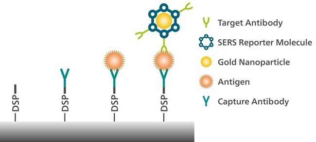
Figure 2. Detection of an antigen associated with pancreatic cancer is possible with surface-enhanced Raman spectroscopy (SERS). Image Credit: Metrohm AG
Rapid, point-of-care (POC) assay for femtogram-level detection of COVID-19
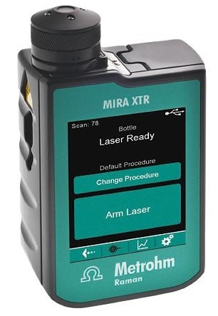
Figure 3. MIRA XTR, a handheld Raman spectrometer from Metrohm. Image Credit: Metrohm AG
Researchers at the University of Wyoming used an alternate ELISA format to identify antigen biomarkers linked with COVID-19 infection.4 This investigation utilized a magnetic nanoparticle-supported assay to concentrate the target biomarker in solution for subsequent SERS detection with MIRA XTR (Figure 3).
It outperformed commercial lateral flow tests in terms of sensitivity, compatibility with solvent and saliva samples, adaptability to new viral variations, and POC detection of COVID-19.
Lateral flow immunoassays produce rapid results, however they only detect at the nanogram level and have limited quantification. In comparison, the SERS-based ELISA is sensitive to femtogram amounts of antigen and provides quick results at the point of care using a commercial portable Raman device.
Multiplex immunophenotyping of blood and breast cancer cells with Raman spectroscopy
Another study used MIRA DS to evaluate a portable SERS-based ELISA for immunophenotyping of various types of red blood and breast cancer cell surfaces.5 Differentiating healthy and infected cells, as well as detecting multiple biotargets in a single sample, can aid in the informed treatment of various types of breast cancer.

Image Credit: Adobe Stock
This test was more specific, sensitive, and repeatable for immunophenotyping in diverse cell types using a smaller analysis sample volume than traditional multiplex immunoassays, and it was less labor-intensive and technically easier to execute.
The authors appreciated MIRA’s Orbital Raster Scan for boosting assay sensitivity by interrogating a larger area and taking spatially averaged values.
Conventional multiplexed flow assays can be restrained by the availability of different colored stains and the interpretation of the data.
They are also associated with a high lab footprint. In comparison, this approach based on portable Raman spectroscopy has the potential to produce precise POC results with a short sample-to-result time, multiplexing capacity, and highly compact equipment.
Easy detection of enzymes with the electrochemical-SERS effect
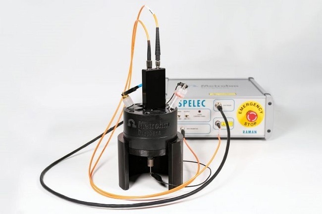
Figure 4. The SPELECRAMAN638 instrument from Metrohm performs spectroelectrochemical Raman measurements using a 638 nm laser. Image Credit: Metrohm AG
Metrohm has reported a rather different SERS approach for the characterization of biological molecules.6
Electrochemical SERS (EC-SERS) enables two experiments at once: electrochemical activation of SERS features of silver electrodes (Ag SPEs), followed by spectroscopic identification of the sample (with SPELECRAMAN638, Figure 4).
The SERS substrate is created in situ using silver electrodes (screen-printed and traditional).
This is achieved in the presence of the analyte while under continuous Raman interrogation to optimize the detection of SERS-active species. The 638 nm excitation produces a strong SERS effect while reducing the danger of sample damage and fluorescence.
Determining the structure of enzymes (and their involvement in illness), such as aldehyde dehydrogenase (ALDH), aids in disease understanding. With EC-SERS, application scientists discovered previously unreported fingerprint Raman bands of ALDH in solution.
Likewise, the redox states of cytochrome c offer information regarding electron transport across cell membranes.7 Cytochrome c undergoes oxidation and conformational changes during the EC experiment, and these redox states have distinct SERS spectra.
Conclusion
Raman technology is increasingly utilized in innovative ways due to its numerous benefits, such as high sensitivity, capability for trace detection, compact size, and ability to deliver rapid results.
Researchers worldwide are applying this technology to identify malignant tissues, detect disease biomarkers, and identify pathogens responsible for illnesses. The outcomes of these applications are both fascinating and promising.
References
- Lindtner, R. A.; Wurm, A.; Pirchner, E.; et al. Enhancing Bone Infection Diagnosis with Raman Handheld Spectroscopy: Pathogen Discrimination and Diagnostic Potential. IJMS 2023, 25 (1), 541. DOI:10.3390/ijms25010541
- Thomas, R.; Bakeev, K.; Claybourn, M.; Chimenti, R. The Use of Raman Spectroscopy in Cancer Diagnostics. Spectroscopy 2013, 28 (9), 36–43.
- Goonetilleke, K. S.; Siriwardena, A. K. Systematic Review of Carbohydrate Antigen (CA 19-9) as a Biochemical Marker in the Diagnosis of Pancreatic Cancer. Eur J Surg Oncol 2007, 33 (3), 266–270. DOI: 10.1016/j.ejso.2006.10.004
- Antoine, D.; Mohammadi, M.; Vitt, M.; et al. Rapid, Point-of-Care ScFv-SERS Assay for Femtogram Level Detection of SARS-CoV-2. ACS Sens. 2022, 7 (3), 866–873. DOI:10.1021/acssensors.1c02664
- Wang, J.; Koo, K. M.; Trau, M. Tetraplex Immunophenotyping of Cell Surface Proteomes via Synthesized Plasmonic Nanotags and Portable Raman Spectroscopy. Anal. Chem. 2022, 94 (43), 14906–14916. DOI: 10.1021/acs.analchem.2c02262
- Metrohm AG. Easy Detection of Enzymes with the Electrochemical-SERS Effect; AN-RA-008; Metrohm AG: Herisau, Switzerland, 2023.
- Brazhe, N. A.; Evlyukhin, A. B.; Goodilin, E. A.; et al. Probing Cytochrome c in Living Mitochondria with Surface-Enhanced Raman Spectroscopy. Sci Rep 2015, 5 (1), 13793. DOI:10.1038/srep13793
About Metrohm AG
Metrohm is one of the world’s most trusted manufacturers of high-precision instruments for chemical analysis. Metrohm was founded in 1943 by engineer Bertold Suhner in Herisau, Switzerland. Today, Metrohm is represented in 120 countries by subsidiaries and exclusive distributors. The global Metrohm Group also includes the Dutch companies Metrohm Applikon and Metrohm Autolab, manufacturers of online analyzers and instruments for electrochemical research, respectively. Recently, the Metrohm Group was joined by Metrohm Raman, a leading manufacturer of handheld Raman spectrometers.
Metrohm is the global market leader in analytical instruments for titration. Instruments for ion chromatography, voltammetry, conductivity, and stability measurement make the Metrohm portfolio for ion analysis complete. Instruments for Near-infrared and Raman spectroscopy are another, strongly growing segment of the Metrohm portfolio.
Metrohm is a problem solver, both in the laboratory and within the industrial process. To this end, the company offers their customers complete solutions, including dedicated analytical instrumentation as well as comprehensive application know-how. More than 30% of the company’s employees at the Metrohm international headquarters in Herisau work in R&D.
Metrohm has been owned 100% by the non-profit Metrohm Foundation since 1982. The Metrohm Foundation, which does not exert any influence on the company’s business operations, sponsors gifted students in the natural sciences, supports charitable and philanthropic purposes and, above all, ensures the independence of the company.
Sponsored Content Policy: News-Medical.net publishes articles and related content that may be derived from sources where we have existing commercial relationships, provided such content adds value to the core editorial ethos of News-Medical.Net which is to educate and inform site visitors interested in medical research, science, medical devices and treatments.