The Ultima 2Pplus All-Optical Multiphoton System combines high resolution, deep imaging capability, speed, and operational flexibility, enabling researchers to efficiently perform simultaneous imaging, photostimulation, and electrophysiology.
Since launching the first commercial system capable of concurrent imaging and uncaging in 2003, Ultima multiphoton platforms have become a trusted standard for advanced neuroscience research across the globe.
Built on this established foundation and powered by Prairie View software, the 2Pplus system integrates over ten years of lab insights and direct collaboration with top neuroscientists, resulting in advanced features found only in Ultima systems.
Key advantages of Ultima 2Pplus
- Industry-leading field of view for multiphoton imaging
- High-performance light collection and detection
- Simultaneous imaging and photoactivation
The neuroscience community has long desired microscopes that support both 3D, high-speed photoactivation and full-field, deep imaging. Bruker’s novel Ultima 2Pplus promises a single-tool solution, providing a new all-optical resource for quantitative investigation."
Dr. Adam Packer, Oxford University
Take the next step
How multiphoton imaging works
In confocal microscopy, a fluorophore absorbs a single visible-light photon, triggering the emission of a fluorescent photon. Two-photon microscopy differs in that the fluorophore simultaneously absorbs two photons, each carrying half the energy (or twice the wavelength) of a single-photon excitation source.
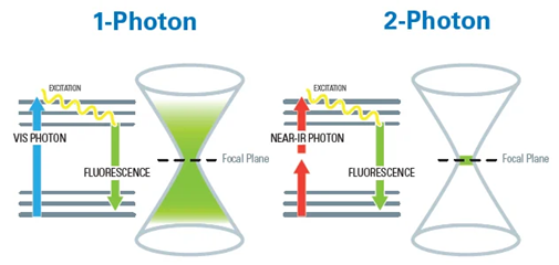
Image Credit: Bruker Nano Surfaces and Metrology
To achieve this effect, two-photon excitation relies on high-intensity, pulsed lasers that deliver energy within ultrashort bursts. These pulses are tightly focused to a specific point within the sample, where the photon density is high enough to induce fluorescence.
Because two-photon absorption is highly localized, fluorescence is generated only at the focal point. In regions with lower photon density, absorption does not occur, significantly reducing background noise and minimizing out-of-focus signal.
Technology for evolving research needs
The Ultima 2Pplus features an optimized optical train that maintains high performance across the full width of the imaging field. Its extended-clearance stage supports imaging of large animal models.
An optically corrected, decoupled electrically tunable focusing module enables simultaneous holographic stimulation and 3D imaging, making the system especially well-suited for advanced neuroscience studies in awake animals.
Looking ahead, the Ultima 2Pplus also supports long-wavelength 3-photon imaging (up to 1700 nm), enabling deep-tissue investigation in living specimens.
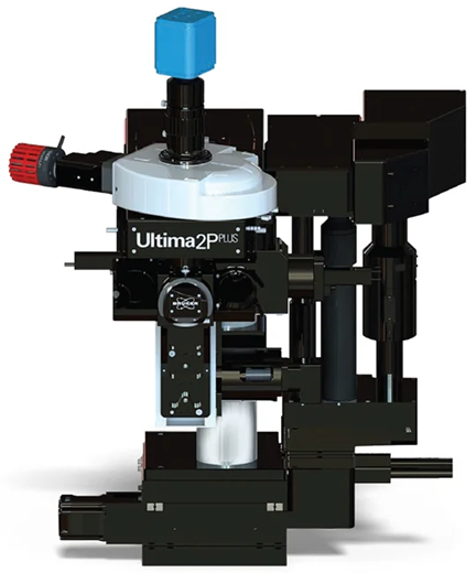
Image Credit: Bruker Nano Surfaces and Metrology
See more, learn more
Largest field of view and superior field uniformity
The Ultima 2Pplus offers a field of view more than 50 % larger, supporting optogenetics experiments that involve simultaneous imaging and stimulation of distant regions within a sample. A newly engineered optical system preserves both spatial and temporal resolution across the entire field, enabling researchers to maintain their existing experimental setups without modification.
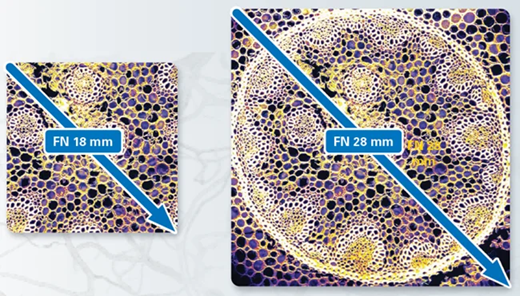
Field of view on Ultima 2Pplus is FN 28 mm in comparison to industry standard of FN 18 mm. Image Credit: Bruker Nano Surfaces and Metrology
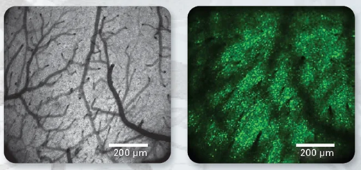
(Left) Exemplary two-photon imaging of cortical surface through a cranial glass window on Ultima 2Pplus. (Right) GCaMP6s expression in layer 5A neurons of mouse visual cortex at a depth of ~400 μm below the pia. Both acquired with Nikon 16x 0.8 NA objective at 920 nm. Scale 200 μm. Data courtesy of Dustin Herrmann, Mehmet Fisek, Michael Häusser’s lab, UCL, London. Image Credit: Bruker Nano Surfaces and Metrology
Look deeper
High-sensitivity deep imaging
The Ultima 2Pplus is engineered to capture nearly all scattered photons from dim, thick, or opaque biological samples. Its high collection efficiency is enabled by 2-inch or larger optics throughout the emission path, paired with sensitive detectors positioned close to the sample.
While two-photon microscopy is widely established, emerging three-photon techniques open new possibilities for deep imaging. Because longer infrared wavelengths scatter less, three-photon excitation may allow imaging several millimeters into brain tissue. Ultima 2Pplus already supports this capability, extending the system’s potential for advanced research.
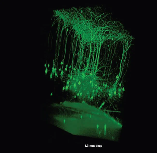
3D-view of pyramidal cells in layer 5. Data collected in one-month-old transgenic Thy1-YFP-H line mouse through a glass-covered cranial window. Data courtesy of Guang Yang, School of Medicine, New York University, New York. Image Credit: Bruker Nano Surfaces and Metrology

3D-view of volumetric stack recorded using the electrically tunable focusing module. Layer 5B neurons in mouse visual cortex in vivo, labelled with tdTomato. Scale bar 100 μm. Data courtesy of Lisa Bauer, Dustin Herrmann, Mehmet Fisek, Michael Häusser’s lab, UCL, London. Image Credit: Bruker Nano Surfaces and Metrology
Acquire even more data
Perfected photostimulation and imaging
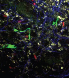
The Ultima 2Pplus supports advanced optogenetics experiments through Bruker’s Neurolight 3D™ Spatial Light Modulator. This module uses computer-generated holograms to simultaneously stimulate multiple targets across different focal planes.
During 3D holographic protocols, targets at varying depths can be imaged using a custom two-photon module with an electrically tunable focusing system. This enables independent Z-positioning of the imaging plane relative to the photostimulation plane.
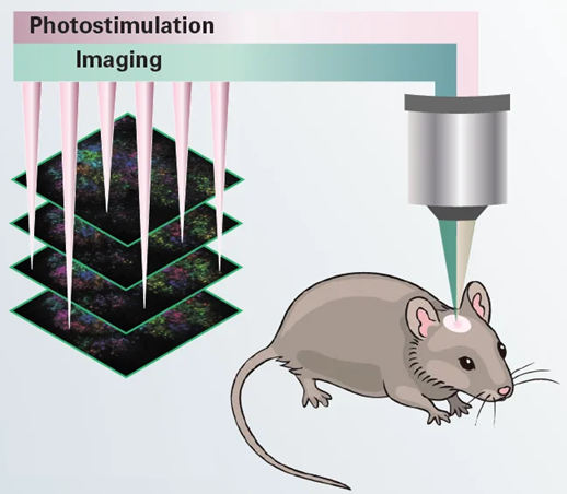
Illustration of 3D simultaneous photostimulation and imaging in vivo experiment. Image Credit: Bruker Nano Surfaces and Metrology
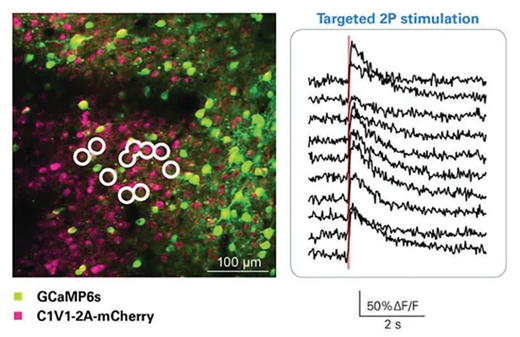
Optical stimulation of circled cells with Neuralight 3D. 3D hologram of points was created which was then spirally scanned over neuronal cell bodies. Data Courtesy of Adam Packer, Lloyd Russell, Henry Dalgleish, Michael Häusser’s lab, UCL, London. Image Credit: Bruker Nano Surfaces and Metrology
Expand the capabilities
Unmatched options for extensibility
One of the Ultima system’s core strengths is its flexibility. A wide range of optional modules allows researchers to tailor the system to meet specific experimental needs:
- Photostimulation Path Module – Enables simultaneous photostimulation or uncaging alongside imaging.
- LED Module – Supports full-field optogenetics applications.
- Resonant Scanner Module – Delivers rapid data acquisition at 30 Hz with 512×512 resolution.
- Rotating Nosepiece Module – Provides off-axis imaging to access regions beyond the vertical axis. Ideal for reducing light scatter in tissue with highly aligned cellular structures. A motorized version is available with both manual and software control.
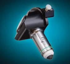
Rotating nosepiece. Image Credit: Bruker Nano Surfaces and Metrology
- Dual Wavelength Imaging Module – Allows two lasers to be directed into the imaging path, enabling multi-probe imaging without retuning. Scanning can occur either simultaneously or in a line-interlaced mode.
Dual wavelength imaging in lymph node explant. Image Credit: Bruker Nano Surfaces and Metrology
- Moving Scope Stage Module – Offers controlled repositioning of the microscope around the sample.
- Moving Sample Stage Module – Includes a low-profile, motorized specimen stage. When paired with the Bridge Stage, the system converts easily into a slice rig.
- Objective Z-Piezo Stage Module – Supports fast volumetric imaging. Fully software-controlled and compatible with resonant scanning synchronization.
- Remote ETL Module – Features an optically corrected electro-tunable lens with a 450 µm range for flexible focus control.
- FLIM Module – Supports Fluorescence and Phosphorescence Lifetime Imaging with dedicated electronics and acquisition software.
- Substage Detectors Module – Optimized for in vitro use, enhancing fluorescence detection in low-signal samples and enabling SHG forward signal capture.
- Dodt Gradient Contrast Module – Improves image contrast in unstained samples, such as brain slices, during electrophysiological recordings.
Ultima 2Pplus specifications scanning
Source: Bruker Nano Surfaces and Metrology
| . |
. |
| Scanning Method |
Matched pair of 6 mm Cambridge galvanometers with raster and spiral scanning capabilities |
| Field of View |
~1.375 mm x 1.375 mm with 16x objective (≤28 mm FN) |
| Scan Speed |
Raster scan: 1.65 fps at 512 x 512, >12 fps at 64; Spiral scan: 6 fps at 512 x 512, ~30 fps at 64 x 64 |
| Scan Customization |
User-definable straight, freehand, and circular (infinite) linescan with included software; User-definable pixels/line and lines/scan from 1 to 2048; ≤120x scan zoom; 360° of scan rotation; Point scan |
| Uncaging Option |
Second set of matched 3 mm or 6 mm Cambridge galvanometers in same scan head provide high-precision visible or multiphoton laser sample photomanipulation |
High-Speed
Imaging Option |
8 kHz resonant galvanometer; ~30 fps at 512 x 512, >1300 fps at 512 x 8 region of interest |
| DETECTORS |
Reflected
Non-Descanned |
1 to 4 hand-picked Hamamatsu Multi-Alkali PMTs; Upgradeable to high-sensitivity Hamamatsu GaAsP PMTs |
Transmitted
Non-Descanned |
1 or 2 hand-picked Hamamatsu Multi-Alkali PMTs; Upgradeable to high-sensitivity Hamamatsu GaAsP PMTs |
| Dodt |
Single Hamamatsu PMT for DIC-like image collection |
| Transmitted |
Single Hamamatsu PMT for transmitted light image collection |
| Camera |
Standard C-mount camera port built into scan head |
| OPTICAL INPUTS |
| Multiphoton Laser |
Optimized for multiphoton laser input from 690 to 1700 nm |
| Epifluorescence |
High-Powered LED in epifluorescence turret |
| Visible Laser |
Fibered laser inputs for visible laser introduction |
| Ultima Laser Rating |
Class 1 (Contains Class 3b and Class 4 lasers) with light box |
| Helios Laser Rating |
Class 3b |
| LED |
Full-field photoactivation with LED module |
| MOTOR CONTROL |
| X,Y Stage |
Variable height with ~15 cm X and ~7.5 cm Y movement and ~0.3 μm step size |
| X,Y Microscope Base |
Fine and coarse-movement platform for scope with ~35 mm travel and 0.1 μm step size |
| Z-Focus |
Range of 30 mm with ~0.2 μm step size |
| Z-Piezo |
Travel range ≤1000 μm with 0.05 μm step size |
| Orbital Nosepiece |
Motorized control of objective angle and rotation |
| SOFTWARE |
| Prairie View |
Turnkey intuitive and customizable operation for imaging |
| Z-Series |
Easy creation of depth stacks with user-customizable slice number, step size and laser power |
| T-Series |
Easy creation of complex series involving Z-Series and triggered images |
| Stage Montage |
Atlas Imaging simplifies setup and optimizes acquisition of 2D and 3D stage montages |
| Peripherals Integration |
Wavelength and power control available for multiphoton and visible laser launches |
| Photoactivation |
User-defined points and regions with synchronized laser modulation |
| Regions of Interest |
User-defined regions for faster scanning capabilities |
| Brightness |
Intensity mapping and plotting for user-defined regions over time |
| Voltage Inputs/Outputs |
Signal inputs and outputs for electrophysiological experiments, stimulus control, and synchronization with external devices |