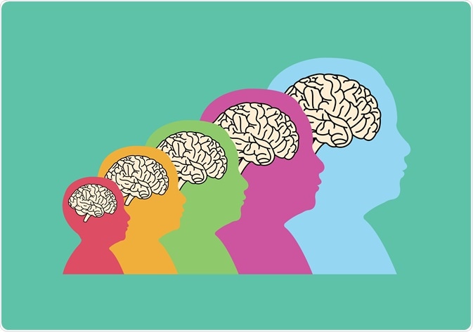
Neurodevelopment is an incredibly complex, but tightly controlled process governed by the sequential action of various genes. Neurodevelopment continues well after birth into early adolescence. Impairments to neurodevelopment at any stage can give rise to neurodevelopmental disorders.

Neurodevelopment. Image Credit: J.K2507/Shutterstock.com
Neural Tube & Early Brain Structures
At the very beginning of embryogenesis, 3 germ layers develop; the ectoderm, mesoderm, and endoderm. The ectoderm gives rise to the brain and skin (epidermis) and lies on top of the developing embryo’s mesoderm which gives rise to skeletal muscle, blood vessels, dermis, and connective tissues. The endoderm is the bottom layer that gives rise to the gut, respiratory epithelia, and many of the body’s other organs.
The brain starts as a simple sheet of ectodermal cells called the neural plate around the end of the 2nd week of embryogenesis (on day 13; E13). Immediately below the neural plate, a group of mesodermal cells form the notochord – present in all vertebrates. The notochord is what initially causes the folding in of the neural plate (neural groove) causing the ends of the neural plate to fuse together forming the neural tube between E20-27. The remaining ectodermal tissue will give rise to epidermal tissues and neural crest cells give rise to many other types of tissue.
The top of the neural tube is covered by a surface ectoderm which has a high expression of BMP & Wnt signaling causing the top of the neural tube to become the roof plate. The bottom of the neural tube is in close proximity to the notochord which has a high expression level of Shh giving rise to the floor plate in the neural tube. This early genetic patterning is what gives rise to the earliest structural organization of the developing brain where the top (dorsal) will give rise to sensory neurons, and the bottom (ventral) will give rise to motor neurons.
The neural tube gives rise to a rapidly developing 3D structure of the early brain that grows almost 10x fold in size from 3-5mm (E27) to 27-31mm by week 8. By this stage, anterior-posterior aspects of the developing brain are well defined by the presence of 3 brain “pouches”: the prosencephalon anteriorly (will give rise to the forebrain), the mesencephalon in the middle which will give rise to the midbrain, and the rhombencephalon posteriorly which will give rise to the hindbrain.
As the embryo continues to develop, the prosencephalon further divides into telencephalon (cerebrum; neocortex) and diencephalon (thalamus/hypothalamus), and the rhombencephalon divides into the metencephalon (pons/cerebellum) and myelencephalon (medulla). These processes are governed by differential gene expression and each of these newly formed areas continues to expand as neurons and glia begin to populate them.
Cortical and Neuronal Development
The neocortex develops from the telencephalon in the developing brain. Anterior-posterior regional patterning is governed by the genetic expression of different transcription factors and regulatory genes. For example, Pax6 and Emx2 are expressed in opposing gradients along with anterior-posterior cortex with Emx2 being highly expressed posterior to medial regions and lowest in anterior regions (in a gradient) whereas Pax6 has the opposite gradient. The highest levels of Emx2 (little Pax6) give rise to the visual cortex (V1). Medium levels of both Emx2 and Pax6 along the middle give rise to the somatosensory cortex (S1). High levels of Pax6 (little Emx2) give rise to the motor cortex (M1).
A key feature of some higher-order mammals (e.g., humans, elephants, dogs, non-human primates, and whales) is the presence of folds (grooves and ridges) on the brain surface called gyri and sulci which massively increase the surface area and volume of the brain. The presence of these on a brain is known as gyrencephalic, whereas the lack of gyri and sulci is lissecephalic – for example, rodent brains. These first begin to emerge around gestation week 8 (GW8) with the first of these being the longitudinal fissure which separates the 2 hemispheres of the brain (finishing at GW22). Other primary sulci form around GW14-26 with secondary and tertiary sulci continuing to form GW30-36.
Neurons begin to form on E42 that will reside in the different regions of the whole brain including within the different layers of the neocortex (discussed below). Before E42, from E25-E42, neuronal progenitor cells symmetrically divide in their masses massively increasing the number of mitotic progenitors (dividing).
From E42, however, these cells begin to divide asymmetrically to give rise to 1 neuron which are post-mitotic cells, and 1 progenitor that can continually divide in this fashion. Neurons begin to migrate out of the proliferative zone (ventricular zone; VZ) into the developing cortex. Cortical neurogenesis is usually completed by around E108 in humans giving rise to the vast majority of adult neurons being present from birth.
Cortical neurogenesis is a very complex process and will be very briefly described in this article for simplicity. This is an incredibly important process that allows the highest order executive and cognitive functions which makes humans “human”. The adult cortex has 6 distinct layers which all have different neuronal populations residing in them. Initially, the first set of developing neurons remain the deepest (except the very first wave of neurons which go right to the periphery) whereas subsequent neurons move outwards in the developing cortex thus forming an “inside-out” pattern.
The very first neurons form and migrate from the ventricular zone (VZ) forming the preplate (PP). The next wave of neurons split the PP into the marginal zone (MZ) and the subplate (SP) which only remain for a short time. Many of these neurons use early scaffolding created by radial glial cells to migrate outwards. The intermediate zone (IZ) between the SZ and SP becomes a mature white matter layer. The cortical plate (CP) develops between the SP and MZ. In the adult brain, none of these embryonic structures remain, but 6 distinct layers remain populated with a plethora of different neuronal types e.g., corticofugal neurons in layers V and VI that project to subcortical areas whereas neurons in layers II-IV are predominantly intracortical neurons which are excitatory glutamatergic neurons.
It is also important to note that gliogenesis and myelination also occur in addition to axonal and synaptic formation – and synaptic changes occur throughout life. Glia cells (https://www.news-medical.net/health/The-Other-Brain-Cells-New-Insights-into-What-Glial-Cells-Do.aspx) (which make up most of the brain) arise from the early multipotent progenitors that give rise to neurons initially, then switching to glia later in embryogenesis (around week 24) after the first set of neurons have formed. An intricate network of blood vessels also begins to form within the brain and the cortical surface as the brain develops.
Impaired Neurodevelopment
Whilst most brains (especially cortices) develop normally according to the tightly controlled sequential manner described above, there may be rare cases where the brain does not develop as planned due to a variety of reasons. These could be due to genetic and environmental factors such as alcohol and drug misuse during pregnancy. Consequently, a variety of different neurodevelopmental disorders can develop with clinical features ranging from barely noticeable (mildest pathology) to severely debilitating both mentally and physically (most severe). Some of these can be apparent from birth or in young infants, others develop later in life such as schizophrenia.
Some key neurodevelopmental disorders include:
- Angelman Syndrome – monogenic postnatal microcephaly caused by loss of function mutations in the maternally inherited UBE3A allele. Angelman syndrome is characterized by severe intellectual disability, absent speech, seizures, developmental delay, and a personality that has a “happy demeanor”.
- Rett Syndrome – monogenic postnatal microcephaly caused by loss of function mutations in the X-linked MECP2 gene (though also associated with CDKL5 & FOXG1). Rett syndrome is characterized by a typical early development, however, this is regressed with the loss of acquired skills, language, intellectual, and motor abilities.
- Autistic Spectrum Disorder (ASD) – a complex genetically and clinically heterogeneous disorder (with causes still not yet fully understood) caused by a variety of genetic interactions/multiple pathways. ASD is characterized by impaired communication and social interactions with some stereotyped behaviors.
- Schizophrenia – another complex disorder with causes still not fully understood though key genetic and environmental triggers are known to be involved. Schizophrenia can develop at any stage in life (not necessarily present from birth unlike the other disorders) and is characterized by positive symptoms (hallucinations and delusions) as well as negative symptoms (blunted affect and lack of motivation).
In summary, the human brain develops in a tightly controlled manner that begins as a simple tube of cells around the 3rd week of gestation. Intricate and complex cellular and molecular signaling pathways give rise to the multitude of complex cell types and cytoarchitecture (including the cortex) of the brain arising from early defined coordinates. The human brain expands massively within the developing cortex by the gyrification of the brain.
Whilst neuronal development is largely completed during embryogenesis, the brain continues to develop well into early adulthood. Abnormal brain development can lead to the onset of neurodevelopmental disorders which are usually present from birth or predispose individuals to neuropsychiatric conditions later in life.
References:
Further Reading
Last Updated: Jul 19, 2021