Background
Principles and challenges
Essential steps and concepts
3D bioprinting techniques used in the creation of bioartificial organs
Successful biomanufacturing of organs/tissues using bioprinting approach
References
Further reading
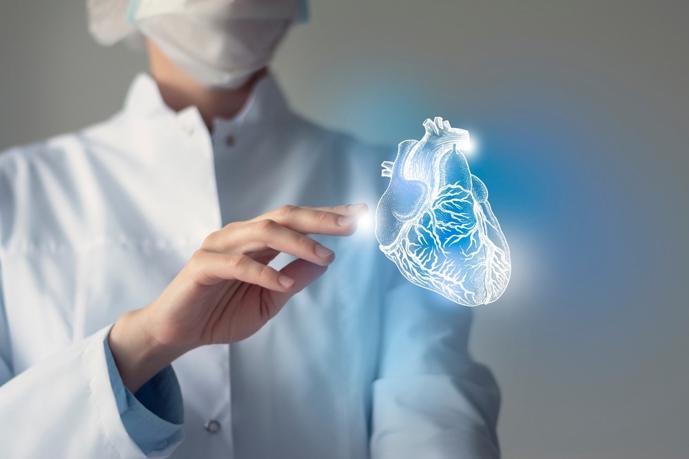
Biomanufacturing of organs/tissues in vitro has been driven by two needs, i.e., organ transplantation and accurate tissue models. Bioprinting, also known as three-dimensional (3D) printing, is a technique used to develop many tissues/organs, such as the liver, skin, and heart. This technique is associated with printing structures using biomaterials, viable cells, and biomolecules. The creation of bioartificial organs has opened new avenues for future organ transplantation programs.

Image Credit: mi_viri/Shutterstock.com
Background
The demand for organ transplants has increased rapidly worldwide. Organ transplants are required due to the increased incidence of vital organ failure and the higher success rate of post-transplant outcomes. Globally, a major organ shortage crisis has emerged due to higher demand and insufficient supply of organs for transplant.
Owing to this crisis, the number of patients on transplant waiting lists is growing rapidly. Thousands of patients are deprived of a better quality of life due to the organ shortage crisis. It has also increased the cost of their medical care, such as dialysis.
Several measures have been implemented to highlight the benefits of organ donation, including increased awareness through educational programs for the public and hospital staff.
Although there is an increase in general willingness to donate organs, it is often not executed by family members due to varied reasons, such as the lack of prompt decision-making ability and emotional state.
3D bioprinting is a new avenue that has shown promise to alleviate the problem of organ shortage and provide patients with a better quality of life.
Principles and challenges
3D bioprinting is an extended application of additive manufacturing (AM) that involves the development of tissues or organs layer-by-layer. This is a versatile technique that uses a bottom-up approach.
Bioprinting aims to mimic the natural cellular microstructure by depositing biomaterial and viable cells precisely. The newly developed organ/tissue by 3D printing looks and performs similarly to the complex natural organ/tissue. 3D bioprinting is based on three fundamental approaches: biomimicry, autonomous self-assembly, and mini-tissue building blocks.
3D bioprinting technology encounters multiple challenges that include gas and nutrient exchange, biocompatibility and biodegradability of the materials used as a substrate, tissue vascularization, shape-fidelity, and restoration of complex functionality of the printed tissue.
Essential steps and concepts
3D bioprinting applies the concept of biomimicry or biomimetics, which is the process of studying nature to stimulate optimal solutions to human problems. In the 3D bioprinting process, the concept of biomimicry is used to choose cells based on an in-depth understanding of the physiology and functions of different cells. These cells would be deposited on a substrate, layer-by-layer, to develop an organ with similar functions and properties.
Autonomous self-assembly is a process that replicates a specific organ/tissue in vitro. This step is similar to embryonic organ development. In a developing tissue, early cellular components synthesize extracellular matrix components and cell signals that promote autonomous cellular organization and patterning of the desirable tissue/organ.
The success of this step is strongly dependent on understanding the mechanism associated with embryonic organogenesis. Furthermore, it is crucial to identify and apply the parameters essential to regulate the embryonic mechanisms.
Materials play a crucial role in cell attachment. Cell type, size, and shape are essential features to regulate cell differentiation and proliferation in a scaffold. Nanoscale features that include ridges and grooves influence cellular attachment and cytoskeletal assembly. A scaffold, a highly porous 3D substrate, is the most essential component of tissue engineering.
Cells grown in a culture medium are transferred to the scaffold. Subsequently, it adheres, proliferates, and produces an extracellular matrix of structural and functional saccharides and proteins that create living tissue. The biological properties of cells are modified and regulated based on the scaffolding material and its internal microstructure.
3D bioprinting techniques used in the creation of bioartificial organs
Different types of 3D bioprinting techniques and strategies are used to develop bioartificial organs. Some of the common techniques are discussed below:
Bio-ink-based 3D bioprinting
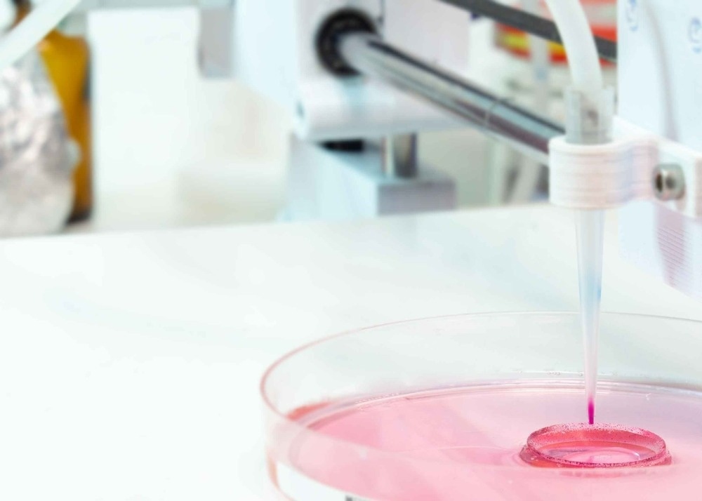
As the name suggests, this 3D printing technique is based on bio-ink, a low-viscosity suspension biomaterial comprising viable cells. Bio-ink can be deposited on a “bio-paper” made of hydrogel substrate, a polymer construct, or a culture dish. This is a non-contact printing approach, which allows printing in a digitally controlled pattern.
Ink-jet printing is conducted following any of the two methods, namely, a continuous manner and drop-on-demand (DOD) printing. The main advantage of bio-ink-based 3D printing is its non-contact nature and cost-effectiveness.
Extrusion-based 3D bioprinting
This method involves direct ink writing (DIW) or pressure-assisted bioprinting methods. This is a material extrusion process where a nozzle continuously extrudes material to create a 3D architecture layer-by-layer. Optimal DIW material possesses rheological properties that promote bioprinting. Typically, polymer resins are blended with fillers, such as nano-clay or silica particles, to achieve suitable rheological properties.
Laser-assisted 3D bioprinting
This method, also known as Laser Induced Forward Transfer, involves using a pulsed laser beam to deposit bio-ink onto the substrate. The use of a laser to deposit cells enables non-contact 3D printing of organs/tissues. The three key elements associated with this strategy are a ribbon coated with bio-ink, a pulsed laser source, and a substrate on which the bio-ink is to be deposited.

Image Credit: luchschenF/Shutterstock.com
Stereolithographic-based 3D bioprinting
This method of 3D printing is based on the height of the design instead of the structure complexity. The stereolithographic bioprinting method utilizes a photo-sensitive heat-curable bio-ink, which is deposited in a plane-by-plane fashion.
The ink must have photocurable moieties since light is used as an agent for cross-linking. Common moieties used for photopolymerization of tissue engineering scaffolds are polyethylene glycol (PEG) and its derivatives.
Successful biomanufacturing of organs/tissues using bioprinting approach
Cardiovascular diseases are the most common cause of death across the world. 3D printing technology has made great progress in enabling heart transplantation, which could be the only option to survive for many patients.
At present, 3D bioprinting technologies have been used to develop heart valves. Laser sintering 3D printers are typically used to create heart valve scaffolds and human umbilical cord blood vessel cells are deposited on the scaffold. Living heart tissues have also been developed using the extrusion printing method. Here, primary cardiomyocytes were obtained from young rats' hearts mixed with fibrin-based bio-inks.
Recently, researchers have developed a cardiac model using 3D printing technology that mimics the realistic feel, mechanical properties, and elasticity of cardiac tissue. This structure could be extremely useful for surgical training applications.
3D bioprinting technology has developed complex liver structures with higher cell density. Recently, scientists at Tsinghua University, China, have biomanufactured different hepatic tissues and organs based on hepatocytes and gelatin-based hydrogels.
Skin is a complex, multi-layered, and the largest organ of the human body. It has been successfully regenerated via 3D bioprinting techniques. A large amount of autologous skin has been manufactured and transplanted in patients with extensive skin defects, including individuals with extensive burns or ulcers.
References
- Tripathi, S. et al. (2023) 3D bioprinting and its innovative approach for biomedical applications. MedComm, 4(1). https://doi.org/10.1002/mco2.194
- Panja, N. et al. (2022) 3D Bioprinting of Human Hollow Organs. AAPS PharmSciTech, 23(5). https://doi.org/10.1208/s12249-022-02279-9
- Song, D. et al. (2021) Progress of 3D Bioprinting in Organ Manufacturing. Polymers, 13(18). https://doi.org/10.3390/polym13183178
- Papaioannou, T. G. et al. (2019) 3D Bioprinting Methods and Techniques: Applications on Artificial Blood Vessel Fabrication. Acta Cardiologica Sinica, 35(3), pp. 284-289. https://doi.org/10.6515/ACS.201905_35(3).20181115A
- Kačarević, Ž. P. et al. (2018) An Introduction to 3D Bioprinting: Possibilities, Challenges and Future Aspects. Materials, 11(11). https://doi.org/10.3390/ma11112199
- de Groot, J. et al. (2015) Decision making on organ donation: the dilemmas of relatives of potential brain-dead donors. BMC Medical Ethics, 16(64). https://doi.org/10.1186/s12910-015-0057-1
- Abouna, G.M. (2008) Organ shortage crisis: problems and possible solutions. Transplantation Proceedings. 40(1), pp. 34-8. doi: 10.1016/j.transproceed.2007.11.067.
Further Reading
Last Updated: Jan 22, 2024