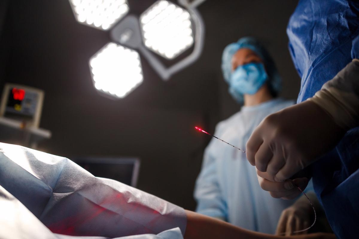
Photonics denotes the science of generation, detection, and manipulation of light waves. Photonic-based methods have contributed significantly to public health in terms of developing rapid, cost-effective, personalized interventions. These methods have many advantages due to the high-speed movement of optical photons and the ability of light waves to penetrate various biological barriers without causing unwanted interactions.

Image Credit: JJ-stockstudio/Shutterstock.com
Photonics in Healthcare
Photonics technologies in healthcare (biophotonics) involve the interaction of light or electromagnetic radiation with living organisms or biological components. This interaction primarily depends on the energy or wavelengths of light and the optical properties of cells. The distribution of the internal refractive index in cells defines their optical properties.
The entire range of light wavelengths is called the electromagnetic spectrum. At both short-wavelength, high-energy gamma rays or x-rays and long-wavelength, low-energy radio waves, the body remains transparent, making it possible to image internal organs and bones non-invasively.
In contrast, infrared and ultraviolet radiations are strongly absorbed by biological tissues. Given this feature, laser lights with infrared/ultraviolet wavelengths are used in a wide variety of biomedical techniques to make tissue incisions, heating or vaporizing specific tissue regions, or seal wounds.
Some bioactive macromolecules can naturally absorb specific light energies or wavelengths in the visible region of the electromagnetic spectrum. The amount of absorbed energy can be used to evaluate the physiological condition of an organ.
Macromolecules that do not absorb light energies can be labeled with specially engineered dyes/biomarkers that can use visible light to highlight specific cell types, such as cancer cells.
Optical Properties of Cells
The refractive index of a cell is associated with its mass distribution. It determines the light propagation in the cell matrix. The refractive index distribution within a cell generates a phase delay for the passing light, measured by quantitative phase imaging.
The refractive index is an inherent structural property of cells. It can be considered a biological marker for cell differentiation or bacterial infection. Cell images obtained from the quantitative phase imaging method can be analyzed to quantify the optical thickness of cells; however, the distribution of refractive index in a cell cannot be obtained from these images unless the shape of the cell is known.
Optical diffraction tomography is another imaging technique used to determine refractive index distribution within cells quantitatively. The two-dimensional phase images obtained from quantitative phase imaging are used for three-dimensional tomographic reconstruction. The tomographic approach is vital for deducing refractive index distribution from optical thickness measured by phase imaging.
Photonics in Disease Diagnosis
Over the last two decades, photonics technologies have been used to rapidly, sensitively, and selectively detect disease-specific biomarkers, metabolites and metabolic biomarkers, pathogens, and disease-specific changes in the composition of cells and tissues, and body fluids.
In optical imaging techniques of the modern era, a laser is used to deliver light in a tissue, and optics or electro-optical sensors are used to detect the diffraction, refraction, scattering and/or absorption of light by the tissue.
Various optical imaging methods are widely used in clinical setups to detect malignancies at the microscopic and macroscopic scale. The most widely used optical imaging methods are endoscopy, Optical coherence tomography (OCT), microscopy, and spectroscopy. Various multimodal approaches that combine all these technologies are also gaining importance. While morphological imaging analyzes structural changes associated with a disease, functional imaging primarily focuses on metabolism, cellular composition, and blood flow changes. Similarly, molecular imaging is used to assess cellular and molecular processes in living organisms.
In ophthalmology, OCT is considered the gold standard for obtaining high-resolution three-dimensional images of the retina. The method is widely used to detect morphological changes in the eye and prevent various eye diseases, including glaucoma and macular degeneration.
Recent advancement in fluorescence endoscopy has made it possible to detect and differentiate small tumors of 1 mm diameter from healthy tissues. Various fluorescent probes, including peptide and nanoparticulate probes that bind to known tumor biomarkers with high specificity, have been developed for early-stage cancer detection. Similarly, label-free methods, including autofluorescence and Raman spectroscopy, have been developed to avoid the expense of fluorescent probe development.
Photonics in Disease Treatment
The delivery of laser radiation through optic fibers is a commonly used method for the targeted treatment of tissues. The treatment outcomes can be modulated by adjusting the wavelength, intensity, and duration of radiation.
In malignancies, diffraction-limited laser light is delivered to small spots. The scattering and absorption of light are analyzed to get information about tumors at the cellular and sub-cellular levels. For incision of tissues, ultra-short laser bursts avoid lengthy surgical procedures, blood loss, and surgery-related pain.
With recent advancements in the field of biophotonics, it is now possible to differentiate malignant tissues from normal tissues and stages of cancer by combining artificial intelligence with photonic technologies.
Many novel biophotonic cancer therapies are under investigation, including endoscopy-assisted surgery, photoablation, photothermal ablation, plasma-induced laser ablation, and photo-disruption.
References
Further Reading
Last Updated: Apr 20, 2022