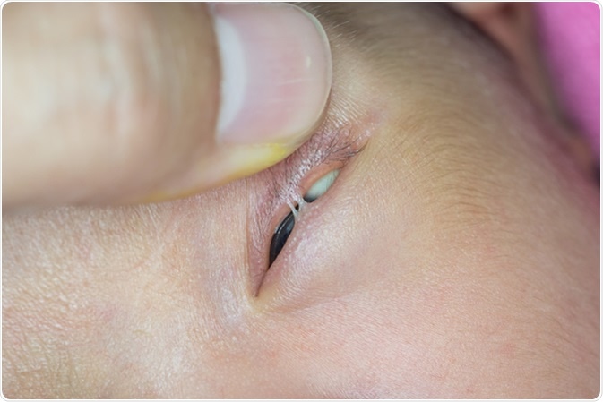
Ankyloblepharon is a term that literally means ‘fused eyelids’. It refers to a condition in which the upper and lower eyelids are fused (or adherent) throughout their thickness by one or multiple connecting strands between the adjacent edges (or ‘ciliary edges’). Both eyelids are anatomically and functionally normal in this condition.
The strands are 1-10 millimeters long and less than half a millimeter in breadth. They arise at the back of the origins of the eyelashes. Moreover, they are composed of a central core of connective tissue with a normal blood supply, surrounded by epithelium.
By pushing apart the eyelids, the opening between them can almost be doubled because the strands are extensible. The extreme ends of the eyelids (the angles of the eyelid opening, called the medial and lateral canthi) are always spared.

Close up of Ankyloblepharon Filiforme Adnatum during eye examination. Image Credit: ARZTSAMUI / Shutterstock
How does ankyloblepharon occur?
In most patients with ankyloblepharon, the condition is present at birth. During embryonic life the eyelids fused until about the fifth month of intrauterine life. At this point they begin to separate, but may remain partially attached until the seventh month.
Ankyloblepharon occurs when the lids fail to separate, either partly or fully, shortening the space between them (the palpebral fissure). This condition can, therefore, be considered as a kind of developmental arrest of the epithelium, while the connective tissue multiplies abnormally fast at the same time. The result is that the developing eyelids are not covered by epithelium at certain places, and therefore remain fused.
What are the types of ankyloblepharon?
Ankyloblepharon may occur in various forms: complete, partial and interrupted. Complete ankyloblepharon is when the eyelids are fused throughout the lid margins. In the partial form, they are joined at one or more points.
Finally, in the interrupted form, which is also called ankyloblepharon filiforme adnatum (AFA), the eyelids show the typical strands. These limit eyelid movement and reduce the width of the opening between the eyelids. AFA has been described in trisomy 18 (also known as Edwards' syndrome).
What are the causes of ankyloblepharon?
Ankyloblepharon may occur either sporadically or as part of a larger syndrome or chromosomal anomaly. For instance, in Hay-Weil’s syndrome of autosomal dominant type, ankyloblepharon occurs along with ectodermal dysplasia and cleft lip or cleft palate.
Familial cases may also be observed, where ankyloblepharon is accompanied by cleft lip and cleft palate.
A third major syndrome is called CHANDS (curly hair-ankyloblepharon-nail dysplasia), and is autosomal recessive in nature.
Other defects such as hydrocephalus, cardiac anomalies, meningocele and various ectodermal syndromes have also been reported. The real importance of this anomaly is that it may be a marker of other congenital abnormalities.
Ankyloblepharon is mostly congenital, but other causes include:
- Chemical injury to the eye
- Traumatic eye injury
- Steven-Johnson syndrome or other disease conditions which lead to scarring
- Eye inflammation caused by e.g. herpes simplex or ulcerative conditions
How is ankyloblepharon treated?
In some cases the bands may be released spontaneously in a matter of few months. Usually, the strands are easily divided under sterile conditions without significant bleeding. If left untreated, the lack of use of the affected eye could lead to amblyopia, or blindness, in the presence of an otherwise normal visual apparatus.
Further Reading
Last Updated: Jun 10, 2023