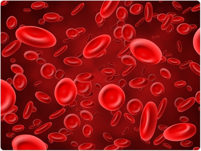
Methemoglobinemia is a potentially life-threatening health condition in which the oxygen-carrying capacity of circulating hemoglobin is significantly reduced.

Image Credit: MicroOne/Shutterstock.com
The biochemistry of methemoglobinemia
The reduction in the oxygen-carrying capacity of hemoglobin (Hb) in methemoglobinemia patients is due to the conversion of some or all of the four iron species being reduced from the ferrous [Fe2+] state to the oxidized ferric [Fe3+] state.
When the iron molecules of Hb are in the oxidized state, they are unable to bind and transport oxygen and carbon dioxide, which can lead to a myriad of health complications.
The biochemistry behind the conversion of Hb into methemoglobin (MHb) can be better understood by reviewing oxidation/reduction reactions. Whereas oxidation involves the removal of electrons from a substrate, the reduction occurs when electrons have been transferred to the substrate.
Since oxidation and reduction reactions always occur together, this combination of reactions is often referred to as “redox” reactions. As Hb is oxidized to MHb, inevitably, oxidation is also occurring in other locations throughout the cell, thereby increasing the likelihood that other cellular enzymes and organelles are damaged.
Causes
Although methemoglobinemia most commonly refers to a condition that arises following exposure to an oxidizing chemical, it can also arise as a result of genetic, chemical, dietary, or even idiopathic etiologies.
Exposure to oxidizing agents
The most common cause of methemoglobinemia occurs following exposure to an oxidizing agent. These chemicals can be categorized as either direct oxidizing agents, which will directly oxidize Hb and form MHb, or indirect oxidizers, which function by reducing oxygen to the free radical O2- or water to H2O2, both of which can oxidize Hb to MHb.
Some of the common oxidizing agents, which can be found in both chemicals and food products, that are associated with causing methemoglobinemia include aniline, benzocaine, dapsone, phenazopyridine (Pyridium), nitrites, nitrates, and naphthalene.
Idiopathic
The second most common cause of MHb is idiopathic and related to systemic acidosis, which will often occur only in young infants younger than 6 months. In these patients, methemoglobinemia will occur secondarily to diarrhea and/or dehydration.
Some factors that increase the susceptibility of small infants to methemoglobinemia include red blood cell (RBC) levels that are low in cytochrome-b5 reductase (CYB5R), as well as more easily oxidized Hb. Since infants often have higher pH levels within their gastrointestinal tract, there is greater support for the production of gram-negative that convert dietary nitrates into nitrites, which are potent MHb inducers.
Dietary
The ingestion of water that contains high levels of nitrates, which is more likely to occur in rural areas where fertilizer runoff can enter public water supplies, can also lead to methemoglobinemia.
Genetic
The congenital forms of methemoglobinemia are typically the result of autosomal recessive defects in the enzyme CYB5R. This type of mutation can lead to a deficiency in either the enzyme CYB5R itself or a deficiency of cytochrome-b5. Typically, infants with this type of genetic defect will present at birth, or shortly thereafter, with cyanosis.
Diagnosis
Since MHb is not capable of transporting oxygen and carbon dioxide, patients with methemoglobinemia are often in a state of hypoxia, which can lead to a wide range of clinical manifestations that range in severity.
Typically, the congenital causes of methemoglobinemia will cause symptoms to present within the first few hours or days of life. Children over the age of 6 months that present with methemoglobinemia will typically develop cyanosis shortly after ingestion.
Like methemoglobinemia, small infants with congenital heart disease will also present with cyanosis; therefore, the distinction between these two disease states can be made on whether the cyanosis resolves with supplemental oxygen.
Notably, methemoglobinemia is associated with the distinguishing feature of elevated PO2 on arterial blood gas analysis, despite having a cyanotic presentation. Comparatively, patients with cyanotic heart disease will have low PO2 and low calculated oxygen saturation.
Regardless of the etiology, the severity of methemoglobinemia symptoms largely depends on the MHb levels, as indicated in Table 1.
|
Methemoglobin Concentration
|
Symptoms
|
|
1.5 g/dL or less
|
None
|
|
1.5 – 3.0 g/dL
|
Cyanosis
|
|
3.0 – 4.5 g/dL
|
Anxiety
Lightheadedness
Headache
Tachycardia
|
|
4.5 – 7.5 g/dL
|
Fatigue
Confusion
Dizziness
Shortness of breath
|
|
7.5 – 10.5 g/dL
|
Coma
Seizures
Arrhythmias
acidosis
|
|
Over 10.5 g/dL
|
Death
|
Table 1: An overview of methemoglobinemia symptoms and their respective MHb concentrations.

Image Credit: Yeexin Richelle/Shutterstock.com
Treatment
The most effective treatment method for methemoglobinemia is the immediate removal of the oxidizing agent, which can be accomplished by tetramethylthionine chloride, which is more commonly referred to as methylene blue.
Upon treatment, nicotinamide adenine dinucleotide phosphate hydrogen (NADPH)-MHb reductase reduces methylene blue to leukomethylene blue through the use of NADPH. Leukomethylene blue then acts as an electron donor to MHb, thereby reducing this harmful component to Hb. Methylene blue is typically administered intravenously within the dose range of 1 to 2 mg/kg over 5 minutes.
In addition to methylene blue treatment, high flow oxygen can also be delivered to methemoglobinemia patients through a non-rebreather mask to increase oxygen delivery to tissues and ultimately increase the natural degradation of MHb in the body.
References and Further Reading
- Ludlow, J. T., Wilkerson, R. G., Sahu, K. K., & Nappe, T. M. (2020). Methemoglobinemia. [Online] Available at https://www.ncbi.nlm.nih.gov/books/NBK537317/. (Accessed 20 August 2020).
- Wright, R. O., Lewander, W. J., & Woolf, A. D. (1999). Methemoglobinemia: Etiology, Pharmacology, and Clinical Management. Annals of Emergency Medicine 34(5); 646-656. doi:10.1016/S0196-0644(99)70167-8.
- Rios, J. D. P., Yong, R., & Calner, P. (2019). Code Blue: Life-threatening Methemoglobinemia. Clinical Practice Cases Emergency Medicine 3(20; 95-99. doi:10.5811/cpcem.2019.3.41794.
- David, S. R., Sawal, N. S., Bin Hamzah, M. N. S. & Rajabalaya, R. (2018). The blood blues: A review on methemoglobinemia. Journal of Pharmacology & Pharmacotherapeutics 9(1); 1-5.
Last Updated: Jul 21, 2023