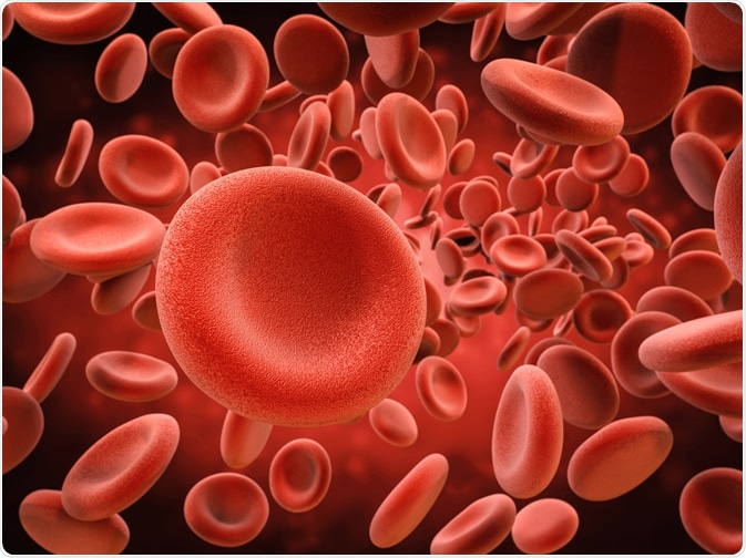
Red blood cell lysis is more commonly known as hemolysis, or sometimes haemolysis.

Image Credit: PhonlamaiPhoto/Shutterstock.com
It refers to the process whereby red blood cells rupture and their contents leak out into the bloodstream. Hemolysis can happen in vivo or in vitro, the first being linked to medical conditions, and the latter being a marked challenge for the medical sector as it hinders clinical testing.
Below we discuss the two scenarios of red blood cell lysis, when it occurs in the body and outside of it, giving an overview of how it occurs and its significance.
Red blood cell lysis in vivo
In vivo hemolysis has numerous causes and is linked with multiple diseases. It is characteristic of a group of conditions known as hemolytic anemias, the causes of which include immune-mediated erythrocyte destruction, such as neonatal isoerythrolysis; incompatible blood transfusion; drugs including penicillin and heparin; fragmentation, such as disseminated intravascular coagulation (DIC), vasculitis, and uremia; hemoparasites, such as Babesia spp.; infectious agents, including Leptospira, Ehrlichia, Clostridium, and equine infectious anemia virus; hypo-osmolality; hypophosphatemia; some chemicals and plants, such as red maple and phenothiazine; and liver failure.
Other causes of hemolysis include artificial heart values, heart-lung bypass machines, pyruvate kinase deficiency, Wiscott-Aldridge Syndrome, HELLP Syndrome, and more. In total, there may be more than 50 causes of in vivo hemolysis. These causes can be classified as immune or nonimmune, hereditary or acquired.
Essentially, hemolysis occurs in vivo when the rate of red blood cell destruction is increased. This leads to hemoglobin being released into the bloodstream. Usually, red blood cells live for around 120 days, and when they die, the spleen removes them from the blood.
The above list of causes each contributes to increasing the rate that red blood cells break down, which can be from mechanical damage or defects in the red blood cells themselves.
The process of in vivo hemolysis usually occurs in the blood vessels and the heart, although it can also occur in the spleen, liver, and bone marrow. It is clinically diagnosed by testing for conditions such as hemoglobinemia (excess of hemoglobin in the blood plasma), hemoglobinuria (excretion of free hemoglobin in the urine), or by testing for decreased serum haptoglobin concentration.
In vivo hemolysis is a serious condition because it plays a role in the pathogenesis of sepsis, causing sufferers to be more likely to develop an infection due to the suppressive effect hemolysis has on the immune system.
Only 2% of blood samples with detectable hemolysis are due to in vivo hemolysis. It is more commonly caused by the skills and practice of medical staff. However, resolving cases of in vivo hemolysis is essential to patient health, although, currently it is difficult to prevent.
Avoiding hemolysis of biological samples
Hemolysis of blood samples before they are tested is a major challenge within the medical industry. It is particularly impactful on emergency departments who need to acquire accurate test results with speed. Hemolysis that occurs in vitro can happen during the collection of the sample, or through the handling of the sample.
It is a significant problem for clinical testing because the lysis of red blood cells can affect the results. The contents that leak from the broken-down cells can directly impact the reading of the results, or they can cause interferences with laboratory analyzers.
The magnitude of the impact is dependent on the type of test being performed and the reagents being used. Tests for potassium, lactate dehydrogenase, and aspartate aminotransferase are particularly affected by hemolysis, where the release of cellular contents into the blood plasma falsely increases the values of the target substances.
Other tests are affected in different ways. When only small levels of hemolysis are detected, the levels are reported along with the test result. When levels of hemolysis are high, the results cannot be relied on, and it calls for a recollection of the sample.
Studies have found that hemolysis is the most common cause for samples to be rejected, with 60% of rejections being attributed to hemolysis. Hemolyzed specimens are five times more frequent than the second most common cause of specimen rejection. This demonstrates the severity of the problem that hemolysis brings to the medical community.
Common causes of in vitro hemolysis are related to the way the blood samples are taken and how they are handled and treated. It is suspected that forcing blood through a too fine needle is the most common reason for samples becoming hemolyzed.
The process can also be caused through the large-bore needle of a syringe into a tube, as well as by shaking the sample too vigorously, or by centrifuging samples before clotting is complete.
Medical professionals are calling for stricter laboratory guidelines to reduce the occurrence of this serious pre-analytical issue. The impact of which is big, costing healthcare systems time and money, and in some cases, leading to incorrect blood test readings that can impact a patient’s level of care.
Sources:
Heireman, L., Van Geel, P., Musger, L., Heylen, E., Uyttenbroeck, W. and Mahieu, B. (2017). Causes, consequences, and management of sample hemolysis in the clinical laboratory. Clinical Biochemistry, 50(18), pp.1317-1322. https://www.ncbi.nlm.nih.gov/pubmed/28947321
Hurley, R. (2007). Anemia and Red Blood Cell Disorders. Immigrant Medicine, pp.611-623. https://www.sciencedirect.com/science/article/pii/B9780323034548500504
Wan Azman, W., Omar, J., Koon, T. and Tuan Ismail, T. (2019). Hemolyzed Specimens: Major Challenge for Identifying and Rejecting Specimens in Clinical Laboratories. Oman Medical Journal, 34(2), pp.94-98. https://www.ncbi.nlm.nih.gov/pmc/articles/PMC6425048/
Further Reading
Last Updated: Jan 30, 2020