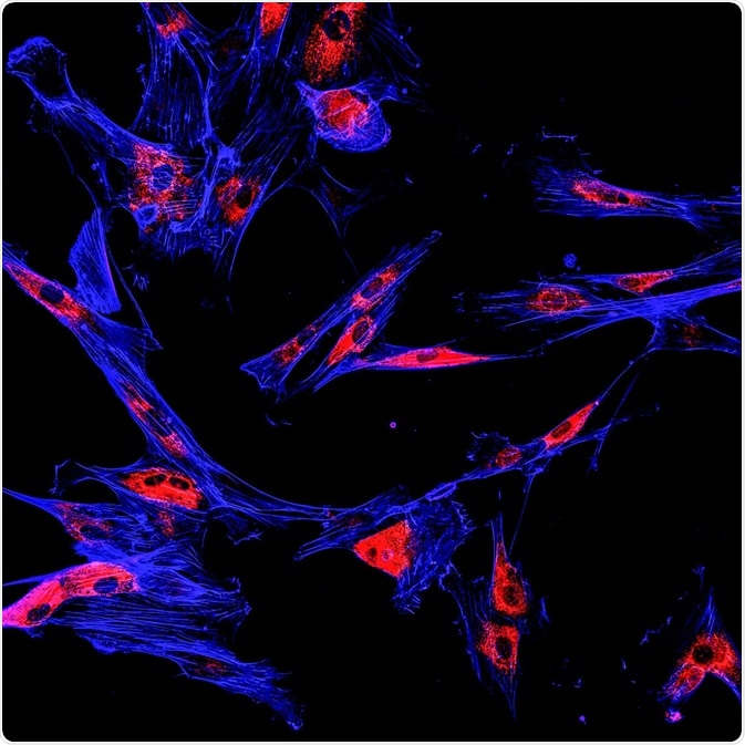
Biophotonics is a technology involving the manipulation of photons in a biological context. It has recently become the mainstay of preclinical drug discovery research.

Credit: Drimafilm/Shutterstock.com
The term biophotonics encompasses the detection, emission, and absorption of photons. The creation, modification, and reflection of light can also be the basis of biophotonic methods.
Common examples of biophotonics studies include fluorescence resonance energy transfer (FRET), biofluorescence, and bioluminescence.
FRET
FRET, also known as Foerster Resonance Energy Transfer, is based on transfer of energy from one fluorophore to another. The emission energy of the first fluorophore, the donor, provides excitation energy for the second fluorophore, the acceptor.
Cyan fluorescent protein (CFP) and yellow fluorescent protein (YFP), are two fluorophores that are commonly used together for FRET. These fluorophores can be engineered into a host cell to study molecular interactions within the cell.
FRET can be used to determine the conformation of proteins or detect protein interactions. It is also a popular method for studying enzyme kinetics.
FRET has wide applications in preclinical drug research. It is a powerful tool for measuring protein-protein interactions, which form the basis of most pre-clinical trials.
Examples of studies carried out using FRET include cell-cell adhesion, cell invasion, membrane matrix metalloproteinase activity, apoptosis, and cell division.
Unlike some comparable methods, the FRET reaction is reversible, meaning the interaction can dissociate. Using this property, a reversible FRET biosensor was developed to monitor inactivation and reactivation kinetics of Src kinase in pancreatic tumors.
The reversibility of the method offers the opportunity to monitor loss of drug-target potency over time, as well as initial drug-target binding and activity.
Biofluorescence and bioluminescence
Many biomolecules are either intrinsically fluorescent or luminescent, or can be designed that way by adding an appropriate chemical group.
This approach can be used to study components of cells or biomarkers, for example. Biomarkers are biomolecules that have been found to indicate a condition of disease or a change in a system.
Biofluorescent molecules allow diseases to be studied using a microscope or an instrument like a spectrophotometer.
Bimolecular fluorescence complementation (BiFC)
BiFC is a technology that allows measurement of protein-protein interactions between separate proteins.
BiFC is being used for studies of transmembrane domain receptor signaling, cellular stress, autophagy, and in vivo protein-protein interactions. It can also be used to study protein degradation.
Fluorescence recovery after photobleaching (FRAP)
FRAP uses fluorescent proteins to mark regions of interest. The fluorescent proteins are then photobleached and the recovery of the fluorescence signal is measured.
FRAP has applicability for membrane dynamics, subcellular diffusion, and the study of chromatin or other processes within the nucleus. FRAP also can be used to study protein-protein interactions.
Photoswitching and photoactivation
Photoswitching and photoactivation are two other mechanisms where fluorescent proteins have be used in preclinical drug studies. These studies have applications for measuring cellular motility, morphology, and intracellular transport.
They have also been used to track tumor cells by repeated imaging of the same region over time, allowing the effects and mode of invasion by tumor cells to be elucidated.
Science Cafe - Biophotonics
Credit: Carleton University/Youtube.com
Further Reading
Last Updated: Feb 26, 2019