Introduction
The early use of flow cytometry in dentistry
What can flow cytometry tell us about our diet?
References
Flow cytometry is a high-throughput cell analysis method that utilizes monochromatic light and specifically angled detectors to infer the cell's size, granularity, and complexity based on the degree of light scattering undertaken.
Cells can also be prepared for fluorescence measurements during flow cytometry by incorporating fluorophores, fluorescent molecules with specific adsorption, and emission wavelengths, allowing multiple non-overlapping markers to be used. Flow cytometry may be utilized in dentistry to examine cells collected from the oral cavity: gums, tongue, uvula, hard and soft palate, etc., including bacterial cells.

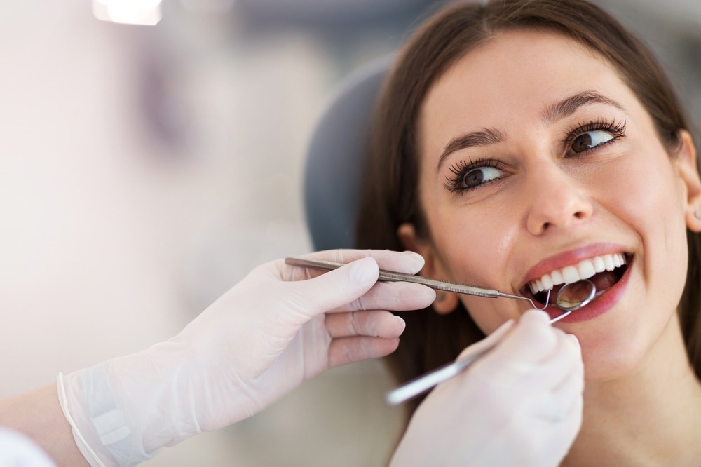
Image Credit: pikselstock/Shutterstock.com
The early use of flow cytometry in dentistry
Mangkornkarn et al. (1991) examined dental pulp tissue in a pioneering study, where flow cytometry allows better reflection of cellular dynamics than previously employed classical methods, where static tissue sections are prepared. The cells were stained with monoclonal antibodies to lymphocyte subpopulations, demonstrating the feasibility of flow cytometry in determining pulpal tissue cellular heterogeneity.
Early steps into the use of fluorescence flow cytometry in oral bacteria identification were undertaken by Barnett et al. (1984), where despite exhibiting distinct light scattering characteristics, Streptococcus mutans and Actinomyces viscosus demonstrated an insufficient difference in cell morphology to enable reliable identification by flow cytometry at the time, with the authors utilizing species-specific immunofluorescent antibodies to achieve separation instead.
Modern flow cytometry can assess several parameters simultaneously and is regularly used in both research and clinical dentistry. In research, flow cytometry is useful in assessing the impact of new antibacterial drugs or materials such as dentin bonding agents (Lapinska et al., 2019), examining the state and cellular composition of oral tissues in disease states, and a wide variety of other specific investigations for which high-throughput multiparametric analysis of cells would be useful. Flow cytometry can be used in the clinic as a diagnostic method for various diseases, where white blood cell count or other indicative biomarkers can be counted and compared.
Several research groups have demonstrated cryopreservation of human dental pulp stem cells and subsequent flow cytometry, making long-term archiving of samples possible (Malekfar et al., 2016). This can be useful in the long-term study of disease states and for intra- and inter-patient comparison studies, particularly in preserving valuable stem cells. Stem cells are regularly identified and categorized in a high throughput manner using flow cytometry and can be collected from the dental pulp in the center of the tooth for use in research and medicine (Bakkar et al., 2017).

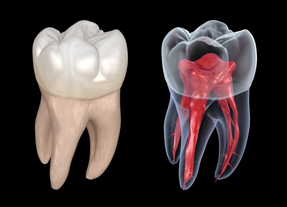
Image Credit: Alex Mit/Shutterstock.com
What can flow cytometry tell us about our diet?
Flow cytometry also has applications in food science-related dentistry. For example, Chang et al. (2012) utilized flow cytometry to examine human gingival fibroblasts under exposure to butyrate, a short chain fatty acid metabolic product of gut microbe digestion of dietary fiber. Interestingly, butyrate inhibited gingival fibroblast growth by cell cycle arrest without inducing apoptosis, producing a toxic effect by producing reactive oxygen species. This may result in prolonging inflammation in periodontal diseases, muddying the already debated health benefits of butyrate (Liu et al., 2018).
A recent study by Onyango et al. (2020) explores the "profoundly different" oral bacteria community and metabolic profile upon exposure to various sugars: sucrose, trehalose, kojibiose, and xylitol, finding that the latter two support fewer tooth decay-associated species. Kojibiose, in particular, was shown to closely maintain the status quo bacterial community composition owing to its low availability to bacterial metabolism.
Kojibiose is a disaccharide with a sweet taste, meaning that fewer sugar calories are required to provide an equal sweetness. It is present in honey but is otherwise difficult to synthesize on an industrial scale, though a recent biochemical approach utilizing enzymes to produce the sugar from a readily manufactured starting material may allow it to be more regularly incorporated into foodstuffs in place of more calorific alternatives (Diez-Municio et al., 2013).
References:
- Mangkornkarn, C., et al. (1991) Flow cytometric analysis of human dental pulp tissue. Journal of Endodontics, 17(2). https://www.sciencedirect.com/science/article/abs/pii/S0099239906816079
- Barnett, J. M, et al. (1984) Automated Immunofluorescent Speciation of Oral Bacteria Using Flow Cytometry. Journal of Dental Research, 63(8). https://journals.sagepub.com/doi/abs/10.1177/00220345840630080501
- Liu, H., et al. (2018) Butyrate: A Double-Edged Sword for Health? Advances in nutrition, 9(1). https://academic.oup.com/advances/article/9/1/21/4849000
- Malekfar, A., et al. (2016) Isolation and Characterization of Human Dental Pulp Stem Cells from Cryopreserved Pulp Tissues Obtained from Teeth with Irreversible Pulpitis. Journal of Endodontics, 42(1). https://www.sciencedirect.com/science/article/pii/S0099239915008997?casa_token=ifRPATJW720AAAAA:JnBrT5lqd6yHi-TX5LtBSWz8Gys6RxJdwbRN8Gv5QKkXbW2PArd47VsDTML8Z3do6lzYu6784Q
- Lapinska, B., et al. (2019) Flow Cytometry Analysis of Antibacterial Effects of Universal Dentin Bonding Agents on Streptococcus mutans. Molecules, 24(3). https://pubmed.ncbi.nlm.nih.gov/30717140/
- Onyango, S. O., et al. (2020) Oral Microbiota Display Profound Differential Metabolic Kinetics and Community Shifts upon Incubation with Sucrose, Trehalose, Kojibiose, and Xylitol. Applied environmental microbiology, 86(16). https://www.ncbi.nlm.nih.gov/pmc/articles/PMC7414948/
- Diez-Municio, M., et al. (2014) A sustainable biotechnological process for the efficient synthesis of kojibiose. Green chemistry, 4. https://pubs.rsc.org/en/content/articlelanding/2014/GC/C3GC42246A
- Bakkar, M., et al. (2017) A Simplified and Systematic Method to Isolate, Culture, and Characterize Multiple Types of Human Dental Stem Cells from a Single Tooth. Adult stem cells. https://link.springer.com/protocol/10.1007/978-1-4939-6756-8_15
Further Reading
Last Updated: Jul 18, 2022