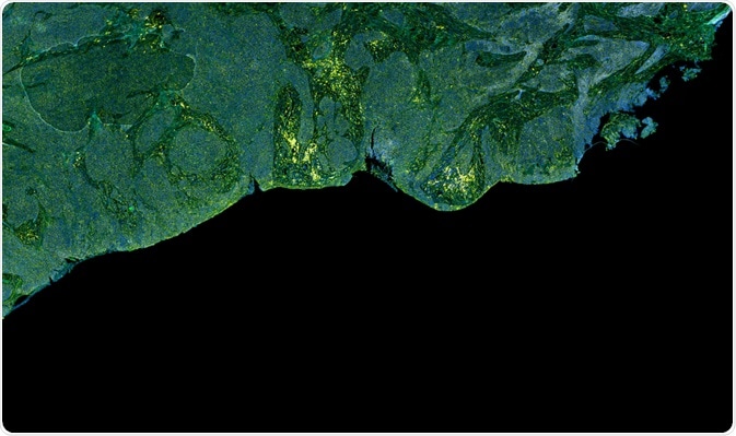
In cancer treatment, surgery remains the best option for removing tumors from affected patients. Successful resection is achieved by the complete removal of tumor cells so as not to cause a resurgence of the cancer. One technique that can aid in recovery rates is fluorescence image-guided surgery (FGS).
 Image Credit: Carl Dupont / Shutterstock.com
The problems with traditional tumor detection and surgery techniques
Typically, margin positivity rates can reach up to 60% and some are considerably less. Cancer surgery relies heavily on visual inspection and physical examination (palpation) by surgeons as well as intraoperative histopathological examination of frozen tumor margins.
However, these methods have their limitations. The naked eye cannot detect certain structures, palpation is limited in sensitivity and is decreasingly being used in favor of robotic techniques, and histopathological analysis of frozen tumors can be time-consuming.
Common preoperative imaging techniques include magnetic resonance imaging (MRI), computerized tomography (CT), and positron emission tomography (PET) but, even though they are effective, there is still a margin of error in surgical margin positivity rate.
It follows that by improving them, a less time-consuming and more effective level of surgical analysis and success can be achieved. Fluorescence image-guided surgery is gaining prominence in this field for this reason.
Fluorescence image-guided surgery – a brief history
The first use of this technology was in 1948 when intravenous fluorescein sodium was used to enhance intracranial neoplasms by G.E. Moore and their team. In recent years FGS has been utilized more widely as new and more effective fluorophores have been developed.
The prominence of FGS as a surgical imaging technique in cancer treatment can be seen by the exponential growth of published articles. In 1990 this was close to zero and grew to 850 in 2018. FGS has achieved a number of clinical successes over the years.
The field of fluorescence image-guided surgery is predicted to grow over the coming years as more clinical research is focused on the techniques. It is a highly effective method that has led to a marked improvement in cancer resection and recovery rates.
How Image-guided Surgery Works
Fluorescence image-guided surgery utilizes two distinct but complementary technologies. These are fluorescence probes and an imaging technique.
The fluorescence probe is usually an organic molecule, for example, a dye or a biomacromolecule (for example, a fluorescent protein) or even a nanomaterial (like a quantum dot). The probe needs to be able to accumulate in the cancerous tissue and not the surrounding healthy tissue.
A limited number of fluorescence probes have been approved by the United States Food and Drug and Administration, even though many do fulfill the aforementioned criteria. The main probes that are approved for clinical purposes are indocyanine green, methylene blue, fluorescein sodium, and 5-Aminolevulinic acid.
Of the aforementioned probes, indocyanine green is the most widely used of the probes. It is a water-soluble near-infrared probe that has been used in 60% of all FGS studies due to its high degree of tissue penetration, high safety index, and low rate of allergic reaction.
The imaging device used has to meet certain critical criteria. Again, the FDA has only approved a limited amount of imaging systems for FGS. The criteria are as follows:
- Ability to overlay white-light and fluorescence images in real-time
- Optimal operation under ambient room lighting conditions
- Nanomolar-level sensitivity to the fluorescence probe
- Quantitative analysis of the image
- Simultaneous detection of multiple fluorophores in the target tissue
- Maximized ergonomic use for open surgery
FGS has been used to treat several cancers including head and neck cancer, ovarian cancer, breast cancer, melanoma, and lung cancer. Some recent research is explained below to highlight the eminent usefulness of this technology.
Using Fluorescence Imaging-guided Surgery to Treat Ovarian Cancer
In 2019, scientists at MIT in conjunction with surgeons and oncologists at Massachusetts General Hospital used FGS to boost the survival rates of patients with early-stage ovarian cancer. Some ovarian cancers are too small to detect with traditional means, even by FGS standards.
Ovarian cancer affects 250,000 patients worldwide, 75% of which are in an advanced stage. Five-year combined survival rates for all stages in the US are 47%. This is only a very slight improvement over a decade ago, where it was typically 38%. This is despite new chemotherapeutic drugs being developed. In breast cancer survival rate is now over 90%.
The research utilized a novel imaging system and approach to FGS to provide real-time imaging of the abdomen with fluorescent labeling of ovarian cancer cells during debulking surgery to remove residual tumor cells which can be too small or hidden from surgeons to ensure proper removal and prevent resurgence of the cancer.
The system developed uses single-walled carbon nanotubes coated with a peptide that binds to SPARC, which is a protein that is overexpressed by ovarian cancer cells. The chemical probe then fluoresces at near-infrared wavelength, even in the presence of the smallest cancer cells, making them easier to image and thus remove.
The team implanted mice with ovarian tumors in the intraperitoneal space and by using the system they could locate and remove tumor cells as small as 0.3mm. No detectable tumors were found after 10 days in the mice. Though tumors did return after 3 weeks, the mice which underwent this technique showed a 40% longer median survival rate than those that did not.
As an intraoperative imaging technique, this approach shows huge potential for the improved survival rate of ovarian cancer sufferers. The researchers plan to do another study in which mice which have undergone this technique are treated with chemotherapy to stop the tumors from spreading. It is hoped that this will show an improved set of results.
Conclusion
Fluorescence image-guided surgery is a cutting-edge scientific technique that is improving the success of cancer surgeries, helping to detect tumors more easily and at an earlier stage, prevent resurgence and proliferation of cancer cells, and improve the survivability of cancer sufferers.
As more novel approaches to this technology are developed in the future, FGS will no doubt play an ongoing and growing part in the treatment of cancer, which is still one of the major causes of mortality and a huge drain on health resources worldwide.
Sources
Nagaya, T et al. (2017) Fluorescence-Guided Surgery Front Oncol. 7: 314 (Accessed 10th May 2020)
https://www.ncbi.nlm.nih.gov/pmc/articles/PMC5743791/
Trafton, A (2019) Imaging system helps surgeons remove tiny ovarian tumors MIT.edu (Website) (Accessed 10th May 2020)
news.mit.edu/2019/imaging-system-surgeons-remove-ovarian-tumors-0424
Zheng, Y et al. (2019) Fluorescence-guided surgery in cancer treatment: current status and future perspectives Ann Transl Med. 7 (Suppl 1): S6 (Accessed 10th May 2020)
https://www.ncbi.nlm.nih.gov/pmc/articles/PMC6462611/
Further Reading
Last Updated: May 28, 2020