BioAFM is seeing increasingly widespread use in biomedical and biological studies due to its extremely high resolution and its capacity to perform experiments with live cells in liquid and under physiologically relevant and ambient conditions.
BioAFM also offers nanometer-resolution surface mapping suitable for an array of electrical and mechanical properties, including stiffness, elasticity, conductivity, and surface potential.
Bruker BioAFM technology empowers life science researchers to explore how these properties influence key cellular functions, including communication, signaling, cell division, differentiation, tumor metastasis, and infection.
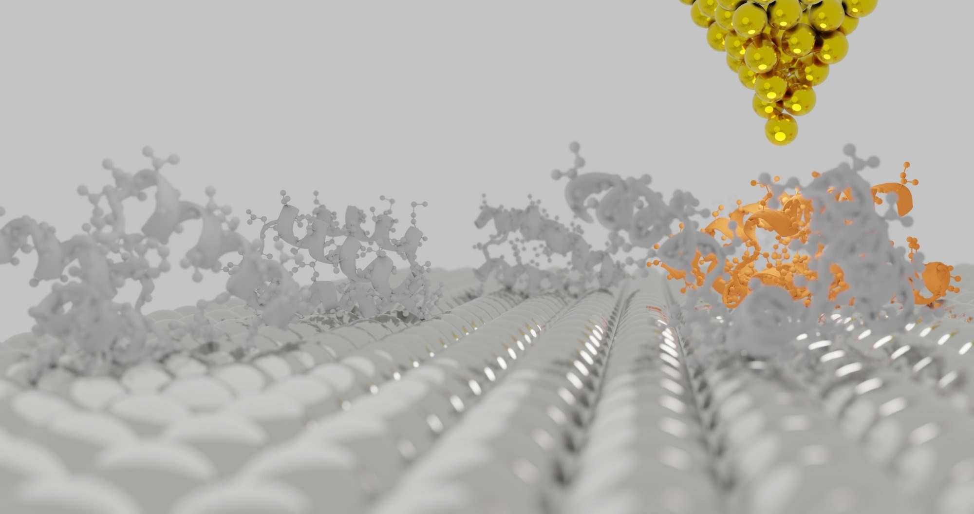
Image Credit: Sanjaya Viraj Bandara/Shutterstock.com
What is a BioAFM?
A BioAFM is a specialized atomic force microscope designed for studying soft matter and biological samples. These instruments are ideal for measuring fragile, soft, and complex samples under near-physiological conditions.
BioAFMs enable the analysis of single molecules, bacteria, living cells, nucleic acids, and tissues without compromising their structural integrity. Their versatility allows for non-invasive measurements of biological samples, including label-free imaging in liquid environments, making them a powerful tool for life science research.
How are BioAFMs adapted for use in biological studies?
BioAFMs feature specialized stages, sample and cantilever holders, and measurement modes, all of which have been specifically designed to accommodate life science samples and experiments.
Another unique feature of Bruker BioAFM instruments is their capacity to be configured with a wide range of optional accessories and advanced modes, expanding measurement capabilities and sample compatibility to accommodate even the most challenging biological and soft matter samples.
Distinct capabilities and features of Bruker BioAFM instruments include:
- High-speed AFM capabilities of up to 50 frames per second facilitate the accurate visualization of molecular dynamics.
- Damage of delicate biological samples is prevented thanks to precise force control.
- Fully integrated correlated analysis is made possible via a comprehensive range of advanced optical techniques.
- Specific data analysis methods are available for soft samples, including contact point imaging.
- A large z range (>100 µm) enables cell adhesion experiments and the measurement of challenging biological samples featuring steep, highly corrugated surfaces.
- Advanced automation features facilitate self-regulating measurement routines designed to maximize throughput and improve accuracy. These features include automated detector alignment, experimental workflow creation, and scanning parameter adjustment.
- An Optical Tiling software feature is available for navigating around large samples.
- Cantilever holders can be cleaned and autoclaved, allowing direct access to biohazards in biosafety laboratories while also enabling sample preparation, loading, experiments, and disposal steps to be performed within the BSL facility.
- Various accessories can be used to investigate a wide range of biological and soft matter samples on different-sized substrates, shapes, and materials (for example, coverslips, Petri dishes, or biochips) and under different environmental conditions (for example, temperature, atmosphere, gas, or pH value).
- The automated, large area, multiparametric characterization of densely packed cell layers and highly corrugated tissue samples can be achieved without the need for microtome cutting.
What are the benefits of using a BioAFM?
Atomic force microscopy delivers three-dimensional images of surface features and topography, and BioAFMs extend, enhance, and optimize this technique to accommodate the specific needs and challenges of biological research.
BioAFM offers a number of distinct advantages over other methods. These include:
- The high spatiotemporal resolution allows users to investigate the morphology and surface structure at the sub-molecular resolution, highlighting interactions in the piconewton range.
- Samples can be studied in liquid and under near-physiological conditions. These capabilities are unique to atomic force microscopy and are ideal for the investigation of living cells in a medium.
- BioAFM’s capacity for the label-free and non-invasive investigation of living cells is ideally suited to use with delicate biological samples.
- BioAFM can be used in conjunction with other advanced optical microscopy techniques, offering correlated measurements and complementary datasets that offer researchers a more comprehensive insight into complex biological processes.
What are the benefits of automated BioAFM measurements?
Bruker’s range of BioAFM instruments boasts specialized software functions and capabilities designed to support the high-performance AFM-based investigation of soft matter and biological samples.
Automated instrument procedures are available, including alignment, operation, and calibration, while automated measurement routines and advanced data analysis capabilities enable the straightforward and streamlined experimental setup and running while improving results’ accuracy and reproducibility.
Advanced automation features include:
Bruker BioAFM instruments incorporate advanced automation and remote control features to streamline complex experiments and enhance research efficiency:
ExperimentPlanner enables users to predefine settings and parameters, allowing for the automated execution of intricate experiments.
ExperimentControl, a browser-based tool, supports remote monitoring of long-term lab experiments from any device. When used alongside ExperimentPlanner, these tools enable self-regulating, long-term experiments that mimic real-life conditions. Researchers can oversee and control their studies remotely, reducing the need for hands-on involvement in lengthy, repetitive procedures.
SmartMapping provides the flexibility to define multiple two-dimensional force maps and preselect regions of interest (ROI) for automated examination. This feature facilitates the systematic study of large sample areas with precision.
Automated large-area, multi-region imaging is achieved through the integration of DirectTiling, DirectOverlay 2, and MultiScan software, extending the optical viewing field. Their seamless optical integration ensures an accurate correlation between AFM and optical data.
Leveraging these advanced features enhances and expands the capabilities of Bruker BioAFM systems to:
- Increase productivity and throughput
- Generate statistically relevant datasets
- Highlight parameter correlation and cross-correlation by automated cycling through relevant parameters
- Enable long-term, unattended, self-regulating experiment series
- Allow the remote monitoring of long-term lab experiments
These types of automated features enable enhanced throughput, standardized batch analysis routines, and statistically relevant datasets—essential considerations in biological research, especially for researchers working in the nanomedical and clinical fields.
Why is it advisable to integrate a BioAFM with an optical/fluorescence microscope?
A significant benefit of Bruker’s BioAFM instrumentation is its ability to be easily combined with advanced optical microscopy techniques such as STED or fluorescence microscopy, enabling complementary datasets and correlated measurements.
Integrating Bruker’s BioAFMs with advanced optical imaging techniques is seamless, thanks to a specialized AFM stage designed for compatibility with most commercially available inverted and confocal optical microscopes.
The AFM head is positioned on the stage, and software such as Bruker’s DirectOverlay feature is then used to colocalize optical and AFM images. This allows for a direct correlation of acquired AFM and optical data.
There is no need to transfer the sample between setups, and the system supports a wide range of camera and detector types.
Easy optical image import, advanced calibration algorithms, and visualization routines enable precise navigation across the sample, allowing for multidimensional characterization within a single experiment.
Compatible techniques include epifluorescence, confocal, phase contrast, and super-resolution microscopy methods such as STED, TIRF, and STORM.
The capacity to acquire real-time, correlative data sets is especially key to the field of life science research. This is because:
- It empowers researchers to study biological samples’ topography using AFM while simultaneously observing fluorescently labeled cellular components.
- It allows the multiparametric observation of in situ dynamics, including protein folding, receptor-ligand interactions, single-molecule protein dynamics, and mechanosensitive signaling pathways.
 Correlative AFM and Advanced Optical Microscopy in Life Science Research eBook
What sample preparation is needed for using BioAFM?
BioAFM is a label-free technique that allows measurements in both air and liquid, making it ideal for studying live cells under near-physiological conditions. Unlike other imaging methods, it does not require a vacuum, nor does it necessitate freezing, drying, coating, or microtome sectioning of samples before measurement.
For optimal results, the sample must adhere to a suitable surface substrate, such as a Petri dish, coverslip, or mica.
When measuring in liquid, the sample should be immersed in an appropriate buffer solution. To ensure high-quality imaging, it is recommended to thoroughly clean the substrate beforehand to eliminate contaminants or artifacts that could interfere with measurements.
Acknowledgments
Produced from materials originally authored by Bruker Nano GmbH.
About Bruker BioAFM
Bruker BioAFM, former JPK Instruments AG, is a leading manufacturer of nano-analytical instruments - particularly based on atomic force microscope (AFM) and optical tweezers systems - for life sciences and soft matter applications.
We combine the highest technical skills with visionary applications. Our work applies nanotechnology in ways to provide solutions to challenges facing researchers in life sciences and soft matter today. Driven by inspiration and ambition, it is our conviction that only the best tools are good enough for the research of life. We are listening with the ear of a scientist in detail to the current challenges of our customers and find individual solutions for individual problems. This is how we understand our business.
Primary Activity
Material Manufacturer
Scanning Probe Technology for Soft Matter and Life Sciences
 Sheffield researchers use JPK's NanoWizard AFM systems to study soft matter, biological systems at molecular scale