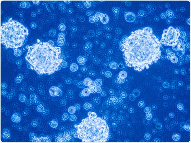
Fluorescence has been used in many different scientific fields including life sciences research and medical applications. The use of fluorescence in surgeries, including those that treat glioblastomas, is improving their success rate.
 Image Credits: Anna Durinikova / Shutterstock.com
What is fluorescence?
Fluorescence is the luminescence caused by the absorption of radiation at one wavelength followed by emission at a wavelength that is usually, but not always, different to that which is absorbed by the material.
Many different materials including gemstones, minerals, and biological molecules (for example, proteins) fluoresce when exposed to incident radiation, usually visible light. The resultant wavelength of emitted light can be used by researchers to identify the target material due to its unique fingerprint. Fluorescence methods target specific fluorophores in the subject material to map their location and arrangement and provide researchers with the information that they need.
What is a glioblastoma?
A glioblastoma is a particularly aggressive form of brain tumor, which accounts for 17% of all tumors discovered in the brains of patients. They are formed from astrocytes and can occur at any age, though they are more common in older people as age is a contributing factor, along with certain genetic predispositions and other factors.
Glioblastomas are progressive, causing increasingly intense headaches, nausea, vomiting, and even seizures as they take hold. They are very difficult to treat and cure. Even with treatment, patients have a median survival rate of 14.6 months when all cases are considered. Up until 2012, cases of aggressive brain tumors increased by an average of 1.2% in the preceding 30 years.
The five-year survival rate for glioblastoma patients is approximately 5%, with 10-year survival rates around 2.6%. If a patient survives for more than 3 years, they are counted as a “long term” survivor.
Scientists working in the field of glioblastoma and brain tumor research are therefore always looking for methods that will improve treatment efficiency and survival rate for patients. In recent years, fluorescence-based methods have shown promise for this purpose.
Fluorescence-guided surgery for glioblastoma treatment
Traditionally, surgical resection for the purpose of diagnosis, relief of effects, and improved survival rate has been the primary treatment for brain tumors along with radiation therapy and chemotherapy. It is imperative in such surgeries to minimize neurologic defects and achieve maximal safe resection of the tumor to improve survival rates without progression of the tumor. Radical resections that may be useful for other types of tumor surgery can lead to increased neurologic morbidity.
The surgeon can utilize several different tools in the operating room including MRI, ultrasound, and neuronavigation to aid in surgeries. However, these can prove to be expensive, time consuming, and ineffective. Specific drawbacks include a high rate of false-positives in MRI and brain-shifts in neuronavigation. Therefore, other methods may prove more useful for the field.
Fluorescence-based methods are proving to have uses in delicate glioblastoma surgeries because they are non-invasive and provide a cheap solution and can provide easy visual identification of tumor cells in real-time without the significant drawbacks of other methods. By labeling tumor cells with fluorescent markers, fluorescence-guided surgery (FGS) can easily differentiate tumor cells from the surrounding brain tissue. When applied to tumor surgery, the procedure is specifically termed a fluorescence-guided resection.
Currently, the only fluorophore that has been approved for fluorescence-guided tumor resection is 5-aminolevulinic acid (5-ALA.) However, use of this fluorophore is not without its problems as protoporphyrin-IX, a metabolic product of 5-ALA that accumulates in target cells, can auto-fluoresce and has limited tissue penetration due to the ability of endogenous fluorophores including heme to absorb its visible-light emissions. Other endogenous fluorophores (e.g. flavin and lipofuscin) present in the brain have excitation/emission spectra that overlap with PpIX.
Near-Infrared fluorescence using indocyanine-green shows improvement in real-time visualization of glioblastoma
A fluorescence-based Second-Window-ICG (SWIG) method that has recently been developed by scientists to overcome these issues is based on near-infrared fluorescence. This method, published online in 2019 in Frontiers in Surgery, uses indocyanine-green, a near-infrared fluorophore that has been commonly used in angiography, being known about since the 1960s. Utilizing the increased endothelial permeability of the tissue around the tumor (peritumoral tissue), the fluorophore is able to accumulate in this tissue.
SWIG has been found to be a highly sensitive method that can specifically target and detect neoplastic tumor tissue in real-time in a wide variety of intercranial pathologies, and has demonstrated utility in craniopharyngiomas, metastases, and pituitary adenomas, as well as gliomas. IGC is a small molecule (<800 daltons) with a half-life of less than 180 seconds.
This proposed method of imaging neoplastic tumor cells expands upon previous studies on ICG since the early 1990s, where ICG boluses were used in a technique to create contrast between the tumor cells and surrounding brain tissue in rat models.
In 2012 a further study was published that demonstrated that using high doses of ICG (7.5mg/kg) 24hrs previous to surgery allowed the fluorophore to accumulate in neoplastic tissue. Enhanced vascular permeability in tumor cells due to defects in their vascular structures and impaired lymphatic drainage systems as well as increased permeability mediators mean that IGC can easily accumulate in these areas and can be visualized up to 24hrs after injection.
The team responsible for this study demonstrated the high sensitivity (up to 98%) of the SWIG method in different clinical trials in 2016 and 2017, amongst other positive advantages of the method.
In conclusion
Neurosurgeons need to be able to precisely differentiate tumors from the surrounding healthy tissue, but this is not always possible with traditional imaging and resection techniques. Fluorescence-guided resection is providing a cutting-edge solution to these problems and research is ongoing into different fluorophores and how effective they might be in aiding glioblastoma surgery and improving survival rates for cancer patients.
Sources
Diaz Valle R et al. (2011) Surgery guided by 5-aminolevulinic fluorescence in glioblastoma: volumetric analysis of extent of resection in single-center experience J Neurooncol. Vol. 102 Issue 1 Pgs. 105-113. Available from: https://www.ncbi.nlm.nih.gov/pubmed/20607351
Glioblastoma – Mayo Clinic. Available from: www.mayoclinic.org/diseases-conditions/glioblastoma/cdc-20350148
Cho, S S. et al. (2019) Indocyanine-Green for Fluorescence-Guided Surgery of Brain Tumors: Evidence, Techniques, and Practical Experience Front Surg. Vol. 6 Issue 11. Available from: https://www.ncbi.nlm.nih.gov/pmc/articles/PMC6422908/
16 Surprising Glioblastoma Multiforme Survival Statistics – Health Funding Research. Available from: healthresearchfunding.org/.../
Further Reading
Last Updated: Jun 2, 2020