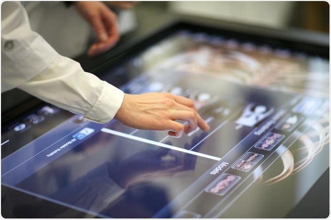
What is digital pathology?
Digital pathology is the practice of sharing digital imagery across laboratories, enabling experts around the world to work together on cases, facilitate the faster development of pharmaceuticals, and to enhance the accuracy of diagnostic techniques.
 Image Credits: Anna Jurkovska / Shutterstock.com
The process involves digitizing the imagery of whole slides of tissue examined under a microscope. These images are then processed, analyzed, stored and managed in an online computer system. As a result, the development of knowledge of numerous diseases and how to treat them is growing.
Its numerous benefits, such as reducing drug research and development timelines, cutting costs, improving tissue analysis, reducing the error rate in diagnosis, enhancing patient outcomes, boosting productivity and facilitating innovation, are helping to accelerate the widespread adoption of the method.
Below we discuss how digital pathology was first established, and how it has developed over the years to get to its current position where it is considered to be setting a new standard of care.
When was digital pathology first established?
Although digital pathology is considered to be a new and emerging technology, the concept has, in fact, been around for decades. The first mention of a methodology resembling digital pathology is over a century old. At this time, scientists were transferring images produced by microscopes onto photographic plates to capture the images and store them.
This enabled the images to be referred back to when studying new cases of the same disease or ailment. It also, for the first time, allowed the images to be shared with experts from other laboratories or those not present at the time of the investigation to gain useful insights, as well as sharing them with those learning the scientific discipline of pathology.
In the late 1960s, the practice of telepathology emerged, where scientists began practicing pathology at a distance, gaining easy access to slide images produced at labs in other locations. At this time, technology had begun to advance to be able to facilitate the sharing of images across locations. In addition to this, NASA’s adoption of telemedicine techniques popularized the idea of the approach as they were seen to successfully monitor the health condition of their astronauts at a distance using telemonitoring systems. Following this, the USA, in particular, began trialing telemedicine techniques on the ground.
However, it has only been during the last twenty years that telepathology has begun to see widespread adoption. Back in the 1990s, whole-slide imaging scanners became commercially available. This was the turning point for digital pathology. Before this, scientists could not produce images of the entire slide sample, requiring them to use their judgment to select regions of interest.
With whole-slide imaging, the entire field is imaged, saving time as human workers are no longer needed to select regions of interest. In addition, as the process has developed it now also saves on errors as it uses AI to automatically select and analyze regions of interest. Removing the element of human error from the process.
The 90s era also saw the rapid development of computer technology in general, meaning that finally, the hardware and software was now available to carry out the data management required by digital pathology. In this time scientists explored how this new technology could be adapted to analyze, visualize, and query data, a process that allowed digital pathology to begin to realize its full potential during the late 2000s.
How has digital pathology developed?
During the last 10-20 years, all industries have undergone a significant shift from analog to digital processes. Medicine is one area that is greatly benefiting from this update in technology. The advent of whole-slide imaging along with this worldwide shift to digital helped to boost the adoption of digital pathology. Whole-slide imaging made it incredibly beneficial, and the shift to digital made it easily accessible.
Since the 1990s vast improvements have been made to the equipment used in digital pathology to digitize the slide images. In addition, huge advancements have been made in the technology in the handling, analyzing, and storing of large amounts of data. This has seen the capabilities and benefits of the method grow over time.
Storage capacities have grown, and AI has been implemented to enhance the speed, accuracy, and efficiency of how the data is handled. This has helped to enhance the widespread adoption of digital pathology.
In addition, these advancements have seen the cost of implementing digital pathology drop considerably. Initially, it cost around $300,000 to acquire and set up the first digital microscope systems. These systems took around 24 hours to scan just one slide. However, today prices had dropped significantly; while costs vary depending on the set up a laboratory has, costs of digital pathology can be as low as $1 per slide.
Furthermore, the advancements in technology have led to higher resolution images that can be imaged in both bright fields and fluorescent conditions. Finally, the software packages now available can also automate and speed up the analysis process.
Overall, these advancements in digital pathology have allowed it to establish itself as a vital tool in modern pathology, having a significant impact on advancing our knowledge of disease and improving the way we treat and diagnose it.
Sources
M. Sergi, C., 2019. Digital Pathology: The Time Is Now to Bridge the Gap between Medicine and Technological Singularity. Interactive Multimedia - Multimedia Production and Digital Storytelling,. www.intechopen.com/.../digital-pathology-the-time-is-now-to-bridge-the-gap-between-medicine-and-technological-singularity
Pantanowitz, L., 2010. Digital images and the future of digital pathology. Journal of Pathology Informatics, 1(1), p.15. https://www.ncbi.nlm.nih.gov/pmc/articles/PMC2941968/
Pantanowitz, L., Sharma, A., Carter, A., Kurc, T., Sussman, A. and Saltz, J., 2018. Twenty years of digital pathology: An overview of the road travelled, what is on the horizon, and the emergence of vendor-neutral archives. Journal of Pathology Informatics, 9(1), p.40. https://www.ncbi.nlm.nih.gov/pmc/articles/PMC6289005/
Further Reading
Last Updated: Jun 5, 2020