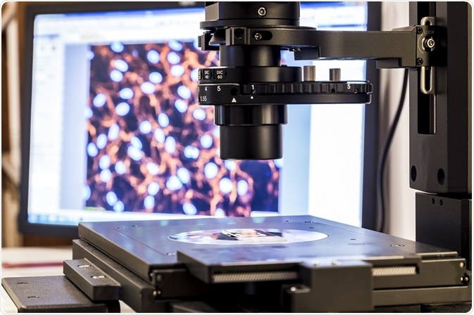
Single-molecule experiments act as a guide in identifying key factors that determine the behavior of biomolecules and their influence on the activities of biomolecules at the single-molecule level.
 dominika zarzycka | Shutterstock
From the initial experiments that are carried out at low temperature, far, or near-field, it is emerging as a potential technique. This technique is crucial for investigating the dynamic heterogeneities that exist in biological systems and complex materials.
In single-molecule experiments, a very small volume, say a femtoliter of the biomolecular solution, is excited and light is collected from it. This results in the single-molecule transition.
Analysis of the concentration regime obtained by the transition over the acquisition time may provide the relevant data that enable to identify the various activities of the particular biomolecule.
Various studies, from protein and polynucleotide pulling to colloidal particle optical trapping, have utilized the single-molecule experiment to successfully measure the fluctuation distribution. The highly simple models of this experiment have facilitated the success of different relevant studies in the past.
Early single-molecule experiments
Numerous advancements achieved by experiments since the 1950s have enabled the application of single-molecule experiments for the study of biomolecules in cells. Instances also abound for using single-molecule experiments in the study of non-biological samples and for low-temperature studies.
The historical relevance of such studies to contemporary single-molecule experiment and the evolution of this new field are discussed below:
Single-molecule experiment in the first generation
In 1956, the first images of single biomolecules such as DNA and proteins, including collagen, were taken with the help of Transmission Electron Microscopy. However, it was difficult to get the direct images of smaller biomolecules, as they possessed constituent atoms that primarily contained carbon atoms of low atomic number.
It is worth mentioning that water is used as the solvent for several biological samples and sample drying also leads to non-native conformations. In 1961, the first indirect study of biomolecules solvated in aqueous solutions was performed.
The fluorescence exhibited by the small droplets (formed due to atomization) that contain low-concentration enzyme and high-concentration substrate was calculated and it was found to be in the order of 0, 1, 2, etc.; this indirectly suggests that the droplets contained 0, 1, 2, etc. number of molecules, respectively.
Era of prosperity for single-molecule experiments
In 1976, Globulin’s single molecules were examined by identifying the relevant molecules in aqueous solution using enormous molecules of fluorescent organic dye.
In 1982, studies of single-lipid molecules were conducted using multiple fluorescent tags that can diffuse through the cell membrane.Intensity centroid localization was used to locate the molecules’ center on a frame-by-frame basis with the aid of photographic papers that are overlaid by tracing papers. This method is now a benchmark in many optical super-resolution and force transduction methods.
In 1988, an experiment was conducted to determine the center of a plastic bead, wherein single kinesin motor was used to rotate the bead to a few nanometers. This localization was performed by digitizing and processing computerized images.
In 1996, the progress attained by the above-mentioned studies was utilized to investigate single-rhodamine-labeled lipids for the first localization of single fluorescent molecule. This study achieved an approximate resolution of 30 nm.
Rise of live and functional cell investigation
In 2000, direct single-molecule imaging of live cells was carried out for the first time, wherein only one fluorescent dye tag was used. The fluorescence microscopy was an inevitable tool for this achievement through single-molecule experiment.
New biocomputational and automated techniques
In recent years, kernel density estimation, Bayesian inference, and edge-preserving filtration were used in carrying out detection in an automated way and completely objective outcomes were gained. In 2013, determination of the spatial dynamics of single molecules was performed utilizing the Bayesian inference.
The single-molecule experiment guides in a greater way in the advancement of “nanotribiology.” A study performed by Pawlak et al. in 2017 shows that when the experimental prerequisites are met, single-molecule experiment is vital for determining the friction-like characteristics of long polymeric chains, single porphyrin molecules, or graphene nanoribbons.
Developments in single-molecule experiment
Although single-molecule detection is a relatively new field, detecting the systems at the single-molecule level is expanding appreciably. These channel studies not only unveil the fact that the single molecule could be investigated, but also contribute several practical, analytical, and theoretical tools.
With reference to DNA repair, understanding the system at the single-molecule level is beneficial for analyzing heterogeneity among the molecular states that take part in the biochemical reaction. In this perspective, the development of this field will have a promising societal impact by offering a new perspective of life at the molecular level.
The single-molecule detection is emerging as a considerable interdisciplinary field that comprises physicists, chemists, and biologists. As this technique is applicable to several fields, it finds varied applications that interest researchers universally in almost all fields of natural sciences and this in turn will benefit the single-molecule experiment with various advancements.
Further Reading
Last Updated: May 24, 2019