Bacterial cell walls; an antibiotic target
For a bacterial cell, the cell-wall is vital in determining the shape of the cell and to maintain its structural integrity. This is a unique feature of bacterial cells, and several antibiotics work by targeting the bacterial cell-wall.

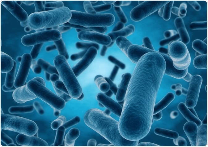
Image Credit: Chiari VFX/Shutterstock.com
The cell-wall needs to be strong enough to counteract the effects of intracellular pressure as well as external pressures, but also needs to have flexibility so that cells can grow and divide; new cell-walls must be created in this process, and to ensure cellular integrity there are mechanisms in place to controls how and when parts of the cell-wall are broken down and re-modeled.
The main component of bacterial cell-walls is peptidoglycan; this is a mesh structure composed of a polysaccharide backbone made of a glycan (poly-[N-acetylglucosamine (GlcNAc)-N-acetylmuramic acid (MurNAc)]), which is cross-linked with short peptide bridges connecting the MurNAc residues.
β-lactam antibiotics, such as penicillin, which are frequently used to treat bacterial infections, target this crosslinking in the peptidoglycan. β-lactam antibiotics prevent this crosslinking from occurring, leading to the accumulation of long glycan strands which are un-crosslinked, eventually leading to bacterial cell death.
Class A penicillin-binding proteins and cell wall defect repair
A study by Vigouroux and co. investigated the role of class A penicillin-binding proteins in E. coli cell-wall shape formation and cell-wall repair. During cell-wall growth, two different enzymatic reactions are required; a transglycosylase reaction and a transpeptidase reaction.
Transglycosylase activity (transglycosylation) extends the glycan, and the transpeptidase reaction (transpeptidation) form the peptide crosslinks between the new glycan strands. In E. coli, this process is carried out by two different “machinery” formed by the combination of various proteins.
One such machinery involves class A penicillin-binding proteins. Class A penicillin-binding proteins are bi-functional, meaning they can carry out transglycosylation as well as transpeptidation. These are activated by LpoA and LpoB, lipoprotein co-factors found in the outer membrane.
In the study, Vigouroux and co. found that class A penicillin-binding proteins inserted new peptidoglycan in areas of the cell-wall which were defective through their intrinsic transglycosylase and transpeptidase activities.
What mechanisms are in place to counteract the effects of β-lactam damage? – example in Pseudomonas aeruginosa
As mentioned above, β-lactam antibiotics disrupt the peptidoglycan and cause the formation of long un-crosslinked glycan strands. A protein in Pseudomonas aeruginosa, Slt, is thought to repair this defect caused by β-lactam antibiotics by a “lytic transglycosylase”, Slt.
In a study by Lee and co., Slt was shown to carry out an exolytic and endolytic reactions, which cleaves glycosidic bonds within the glycan either at the end (exolytic) or the middle (endolytic). The long glycan chain formed by the presence of β-lactam antibiotics is the initial substrate for the endolytic reaction in Slt.
The resulting products then serve as the substrate for the exolytic reaction in Slt, generating the product Ilpa. When IlpA is internalized, a signaling cascade is triggered resulting in the expression of β-lactamases, which confer resistance to β-lactam antibiotics.
Therefore, Slt not only counteracts the cell-wall defects caused by β-lactams by breaking up the long glycans but also triggers resistance to β-lactam antibiotics.
Signaling cascade for cell wall integrity in the yeast Saccharomyces cerevisiae
Yeasts are unicellular fungi and they also have a cell-wall, but this differs from bacterial cell-walls. The cell-wall in Saccharomyces cerevisiae consists of polysaccharides (β-1,3-D-glucan, β-1,6-D-glucan, and chitin) and glycoproteins.
The polysaccharides are thought to form the structure of the cell wall, while the glycoproteins are important in adjusting the permeability of the cell wall. As in bacteria, the cell-wall served to maintain the shape of the cell and to protect the cells from internal and external pressures.
A signaling cascade enables the cell to monitor the integrity of the cell wall. In S. cerevisiae, the small G-protein Rho1 controls the signaling cascade during cell division as well as in response to cell-wall damage.
This signaling cascade not only controls the expression of enzymes involved in cell-wall synthesis and other cell-wall associated genes, but also the polarization of the actin cytoskeleton. One such example is the CWI signaling pathway, which is active during cell division but also can be activated by damage to the cell-wall.
Sources
- Dörr, T. et al. (2019) Editorial: Bacterial Cell Wall Structure and Dynamics. Frontiers in Microbiology https://doi.org/10.3389/fmicb.2019.02051
- Vigouroux, A. et al. (2020) Class-A penicillin-binding proteins do not contribute to cell shape but repair cell-wall defects. eLife DOI: 10.7554/eLife.51998
- Lee, M. et al. (2018) Exolytic and endolytic turnover of peptidoglycan by lytic transglycosylase Slt of Pseudomonas aeruginosa. Proceedings of the National Academy of Sciences of the United States of America https://doi.org/10.1073/pnas.1801298115
- Kollár, R. et al. (1997) Architecture of the Yeast Cell Wall. Journal of Biological Chemistry. DOI: 10.1074/jbc.272.28.17762
Last Updated: Mar 26, 2020