Morphogens are signaling molecules that are vital for cell differentiation. These molecules directly influence cell differentiation, inducing specific cellular responses in relation to their concentration.
While research has found a wealth of evidence to support this method of action, it has proven a challenge to obtain evidence of their direct impact on cells, leaving the list of molecules that are confirmed as fitting the criteria of a morphogen.
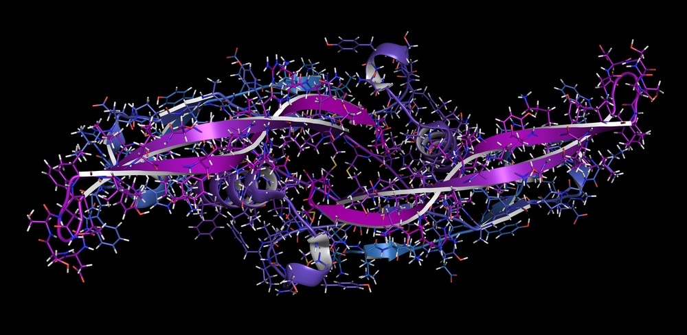
 Bone morphogenetic protein 7 (BMP-7) plays important role in development of bone and cartilage. Image Credit: StudioMolekuul / Shutterstock.com
The Importance of Morphogens
Concentration gradients of morphogens dictate how cells in the body’s tissues differentiate, a vital developmental process. The morphogens originate from a specific region within a tissue, creating a concentration gradient as they diffuse away from this point. Essentially, the gradient codes for how cells differentiate, determining their function and role in the body.
Gaining a deeper understanding of how morphogens work and getting a more accurate view of their signaling pathways is vital to understanding cell development. Additionally, it is essential for the development of new treatments and diagnoses of disorders and illnesses related to morphogen function.
For example, recent research has uncovered that morphogens are a vital part of the neurovascular unit (NVU), a structure essential for the management of the blood-brain barrier (BBB). Given that numerous disorders of the central nervous system (CNS) are linked with impaired functioning of the BBB, research into components essential to its operation is vital to developing new, effective therapies for a wide range of disorders.
Morphogen Signaling Pathways
Distribution of neural subtypes
Given that morphogens work in all kinds of cells and tissue in the body (both human and animal), there is no one single method of action shared between all types of morphogens. Here, we give an overview of one of the most widely studied morphogen signaling pathways, that of the developing vertebrate spinal cord.
The vertebrate spinal cord originates during gastrulation from the neural plate. At the point where the neural plate closes to form the neural tube, patterns begin within the cells of the rostrocaudal (RC) and dorsoventral (DV) axes and the cells begin to differentiate depending on where they are located within the neural tube. Neurons leave the cell cycle and differentiate into specific types depending on their position, a process vital for the assembly of neural circuits.
Research has demonstrated that several families of signaling molecules are involved in controlling patterning and growth of the neural tube. Here, we will talk about the four morphogens considered to be the most crucial: Shh, Wnt, BMP, and FGF. Signaling molecules such as Notch are not discussed due to acting only in short-range and, therefore, not fitting the criteria of a morphogen.
FGF
FGFs signal through receptor tyrosine kinases. They are involved in neural stem cell growth, proliferation control, and CNS patterning and cell specification events during embryonic development. Additionally, FGF signaling (mostly via FGF8 ligands produced by the presomitic mesoderm) is vital to maintaining cells in the caudal stem cell zone of the neural tube.
Wnt
Like FGF morphogen, members of the Wnt family are also responsible for neural proliferation. Currently, studies have highlighted three distinct pathways that transduce Wnt signaling: the canonical, planar cell polarity, and Ca2+. The canonical pathway, for example, involves the stabilization of transcriptional co‐activator b‐catenin before it moves into the nucleus.
Here it interacts with DNA binding proteins from the TCF/Lef family to regulate transcription. Various studies have highlighted the role of the Wnt pathway in cell cycle progression and cell survival in the neural tube, with its role in developing cells of the midbrain and hippocampus being highlighted.
BMP
Morphogens that make up the BMP family of transforming growth factor‐(TGF)β molecules are also found in the dorsal neural tube as well as the overlying dorsal ectoderm. Research is beginning to reveal that the two families (BMP and Wnt) work closely together during neural tube development and their pathways may interact. It is believed that the BMP pathway may have a regulatory effect on wnt signaling.
Shh
The Shh protein acts as a mitogenic and survival factor in the CNS. Whilst BMPs and Wnts are found in the dorsal neural tube, Shh is located in the ventral regions, and its activity is antagonized by members of the BMP family. While it has been shown to have numerous important roles, its most recognized function is that of specifying neuronal subtype identity. In addition, Shh is involved in determining cell fate, as well as encouraging the proliferation and survival of cells.
Overall, the signaling pathways of morphogens are numerous and complex. A mounting body of evidence suggests each family of morphogens has numerous functions and that their signaling pathways interact with those of other morphogen families. As research techniques evolve and we are able to take a closer look at the workings of morphogens our knowledge will expand, enabling us to better understand the developmental process and disease.
Sources
Bijlsma, M., Peppelenbosch, M. and Spek, C., 2006. Hedgehog Morphogen in Cardiovascular Disease. Circulation, 114(18), pp.1985-1991. https://www.ahajournals.org/doi/full/10.1161/circulationaha.106.619213
Sagner, A. and Briscoe, J., 2017. Morphogen interpretation: concentration, time, competence, and signaling dynamics. Wiley Interdisciplinary Reviews: Developmental Biology, 6(4), p.e271. https://www.ncbi.nlm.nih.gov/pmc/articles/PMC5516147/
Smith, J., Hagemann, A., Saka, Y. and Williams, P., 2008. Understanding how morphogens work. Philosophical Transactions of the Royal Society B: Biological Sciences, 363(1495), pp.1387-1392. https://www.ncbi.nlm.nih.gov/pmc/articles/PMC2610127/
Stathopoulos, A. and Iber, D., 2013. Studies of morphogens: keep calm and carry on. Development, 140(20), pp.4119-4124. https://www.ncbi.nlm.nih.gov/pmc/articles/PMC3787753/
Tabata, T., 2004. Morphogens, their identification and regulation. Development, 131(4), pp.703-712. https://dev.biologists.org/content/131/4/703
Wevers, N. and de Vries, H., 2015. Morphogens and blood-brain barrier function in health and disease. Tissue Barriers, 4(1), p.e1090524. https://www.tandfonline.com/doi/full/10.1080/21688370.2015.1090524
Further Reading
Last Updated: Jan 25, 2021