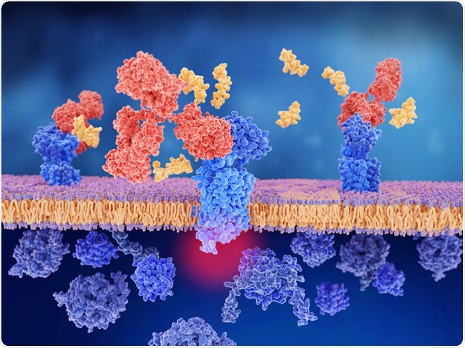
In most clinical applications, the monoclonal antibodies (mAbs) used are of murine origin; however, the small spleen of the mouse is limited in its ability to generate antibodies. Also, the mice used for laboratory experiments are usually inbred in a controlled environment, further restricting their ability to demonstrate diverse immune responses.
 Juan Garter | Shutterstock
Advantages of rabbit mAbs
Apart from having a simpler structure, rabbit mAbs offer higher binding affinity (10 to 100 times more) and specificity than murine mAbs, and they are easier to humanize. These properties of rabbit mAbs make them useful in cases where antigens exhibit weak immunogenicity. Rabbit antibodies can recognize a number of targetable epitopes on humans than rodent antibodies which is pivotal in basic research and in pre-clinical trials.
Production of rabbit mAbs
The rabbit is first injected with the antigen to produce a humoral immune response. From this immunized rabbit, spleen cells are isolated and fused with myeloma cells. After fusion, the hybridoma cells are isolated, screened, and cultured to produce the required antibody.
Different technologies used for rabbit mAb production are hybridoma technology, phage display technology, and other alternative methods.
Hybridoma technology
While rabbit mAbs developed by this technology are valuable reagents for laboratory research purposes and in diagnostic applications, the methodology is difficult.
Phase display technology
This method develops rabbit antibody libraries in single-chain variable format (scFv) as well as in antigen binding fragment (Fab) format. The Fab format has a greater advantage due to its monomeric nature, higher stability, and affinity. scFv formats have the tendency to undergo dimerization, trimerization, and tetramerization.
The phase display method can also be used to engineer single domain antibody formats, such as the N-terminal variable light chain (VL) and N-terminal variable heavy chain (VH) from rabbit mAbs. The smaller sizes of VL and VH make them useful in the recognition of cryptic epitopes.
Alternative methods
Other methods that are used to produce rabbit mAbs and to analyze immune antibody repertoires are high-throughput DNA technologies with mass spectrometry.
Application of Rabbit mAbs
In vitro diagnostic tools
Approximately 11 rabbit mAbs have been approved by the United States Food and Drug administration (USFDA) for in vitro diagnosis in clinics. For instance, rabbit mAbs have been developed to detect the expression of tumor-associated antigens, such as human epidermal growth factor receptor 2 (HER2) in breast cancer, helicobacter pylori infections, circulating tumor cells in prostate cancer, and so on.
Rabbit mAbs are considered superior reagents in immunohistochemistry, western blot analysis, flow cytometry, as well as in the detection of post-transcriptional modification of proteins. An example of a biomarker is the GCET2 rabbit mAb developed by Pan et al. This marker is a sensitive immunohistochemical marker that has demonstrated greater diagnostic sensitivity to detect diffuse large B-cell lymphoma.
Construction of rabbit immune Fab libraries
These libraries are generated to identify mAbs with high affinity and specificity. This is achieved by isolating antibody variable domain repertoires from the B-cell complementary DNA (cDNA) obtained from the rabbit spleen and amplifying it into a Fab repertoire by the polymerase chain reaction (PCR) method.
Applications in oncology
A number of rabbit mAbs are under clinical trials and some are already being used in cancer therapy. For instance, a humanized anti-vascular growth endothelial factor (anti-VEGF) rabbit mAb that is currently under investigation for its effect on metastatic colorectal cancer and other solid tumors. Similarly, another humanized rabbit anti-VEGF mAb is currently under clinical trials for use in age-related macular degeneration.
Rapid detection of Ebola virus
Recently, Wei Y et al developed an ultrasensitive, carbon nanoparticle-labeled pad with rabbit anti-Ebola virus (EBOV)-VP40 immunoglobulin G (IgG) antibody for rapid detection of EBOV. This conjugate pad along with a nitrocellulose pad layered with a monoclonal antibody (McAb, 4B7F9) against EBOV-VP40 and goat anti-rabbit Ig, sample application pad, and absorbent pad were assembled together to form a lateral flow test strip.
The test strip demonstrated strong specificity against EBOV as well as against viruses that were clinically and geographically similar to EBOV, such as yellow fever virus, dengue virus, influenza virus, and Marburg virus.
With distinct antibody repertoires offered by rabbit B cells and the possibility of humanization of rabbit mAbs, therapeutic applications of rabbit mAbs will gain further momentum in the coming years.
Further Reading
Last Updated: Jul 1, 2019