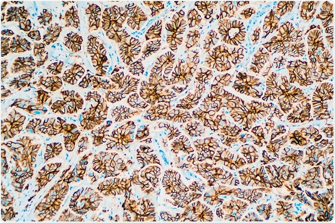
Histone deacetylase (HDAC) antibodies are used to study the process of histone deacetylation, an essential activity that happens within human cells.

Image Credit: David A. Litman/Shutterstock.com
Numerous assays currently exist that rely on the use of histone deacetylase (HDAC) antibodies to explore the proteins present within a biological sample and to view how they interact with other biomolecules, which can uncover information on a wide variety of factors. Assays including DNA pull‐down, ChIP, immunohistochemistry, and Western blot have utilized this kind of antibody. Fields that are benefiting greatly from these methods include cancer research and pharmacology.
The significance of histone deacetylase (HDAC) antibodies
Essential processes that occur within human cells, such as progression of the cell cycle, regulation of transcription, and other developmental activities, rely on the mechanism of acetylation and its reverse process, deacetylation, of lysine residues that reside within DNA’s histones, specifically inside the N-terminal tail domains.
Histone deacetylases (HDACs), alongside histone acetyltransferases (HATs), are vital to the acetylation process. Usually, activities such as condensation of nucleosomes and repression of transcription are linked with the deacetylation process.
During histone deacetylation, histone deacetylase (HDAC) catalyzes the acetyl functional group’s removal from the histones. This can be viewed by the use of methods that exploit histone deacetylase (HDAC) antibodies. The activity of histone deacetylase (HDAC), which leads to a reduction in levels of acetylation, can be measured through the related repression of gene expression that occurs through the condensed state of chromatin.
Research has uncovered numerous histone deacetylase (HDAC) antibodies that fit into four discrete groups. Class one antibodies are found mostly within the nucleus of the cell. Class two, roughly twice the size of class one antibodies, are found in the cytosol and the nucleus. The third class occupies the nucleus and the cytoplasm and are activated by NADH. The final, fourth class has only one antibody allocated to it, HDAC11.
Uses in research
Histone deacetylase (HDAC) antibodies can be used to understand about histone deacetylation within cells. Numerous research methods rely on these antibodies to detect and analyze what proteins exist within biological samples and how they interact. They are currently key to the following research methods:
DNA pull‐down assay:
The DNA pull-down assay is used for detecting protein interactions and can extract protein DNA complexes from a biological sample. The method involves the use of a DNA probe that is isolated by using an antibody against the target protein. The unknown DNA sequence bound by the protein can then be identified by further analysis, usually PCR or Southern blotting. These kinds of studies have, for example, been used to uncover the roles of certain gene expressions in disease, such as in cancer.
ChIP assay:
ChIP, or chromatin immunoprecipitation, is a robust and adaptable assay that is used to view protein-DNA interactions within a cell. It can also identify multiple proteins that are linked with a particular genome simultaneously, as well as to identify the genome associated with a specific protein. Histone deacetylase (HDAC) antibodies have been used in these testing scenarios, specifically, they have been used to uncover protein interactions related to the development of cancer.
Immunohistochemistry:
The process of immunohistochemistry involves identifying antigens (proteins) within the cells of a biological sample, and it is the most common immunostaining technique. It utilizes the fact that antibodies bind with certain antigens within the biological samples. Histone deacetylase (HDAC) antibodies are often used in this assay. Again, this method has been used in the field of cancer research and has been able to identify possible new therapeutic avenues for lymphoid malignancies involving histone deacetylase (HDAC) inhibitors.
Western blotting assay:
Western blotting, also known as immunoblotting, is a commonly used technique for the detection of proteins within biological tissue. It involves separation via electrophoresis, followed by staining with antibodies. Histone deacetylase (HDAC) antibodies are commonly used during the staining process. Staining with these kinds of antibodies has been useful in uncovering the mechanisms underlying tumor growth and progression.
Fields of study
The potential research areas for the use of Histone Deacetylase (HDAC) antibodies are vast. However, there are certain areas for which the use of these antibodies has proven to be more fundamental.
The study of cancer has benefitted greatly from work with Histone Deacetylase (HDAC) antibodies. The above-described assays have been successfully used to uncover the details of various processes that support the initiation and growth of different kinds of cancer. They have been able to highlight the importance of specific genes, and suggest how they influence cancer. Also, pharmacology has relied on the use of this kind of antibody in the development and testing of new pharmacological drugs. They have been used to help identify how drugs will work if they will work for their intended use, and what their efficiency may be.
Sources:
- Gloghini, A., Buglio, D., Khaskhely, N., Georgakis, G., Orlowski, R., Neelapu, S., Carbone, A. and Younes, A. (2009). Expression of histone deacetylases in lymphoma: implication for the development of selective inhibitors. British Journal of Haematology, 147(4), pp.515-525. https://www.ncbi.nlm.nih.gov/pmc/articles/PMC3181219/
- Karch, K., DeNizio, J., Black, B. and Garcia, B. (2013). Identification and interrogation of combinatorial histone modifications. Frontiers in Genetics, 4. https://www.ncbi.nlm.nih.gov/pubmed/24391660
- Murakami, M., Ito, H., Hagiwara, K., Yoshida, K., Sobue, S., Ichihara, M., Takagi, A., Kojima, T., Tanaka, K., Tamiya-Koizumi, K., Kyogashima, M., Suzuki, M., Banno, Y., Nozawa, Y. and Murate, T. (2010). ATRA inhibits ceramide kinase transcription in a human neuroblastoma cell line, SH-SY5Y cells: the role of COUP-TFI. Journal of Neurochemistry, 112(2), pp.511-520. onlinelibrary.wiley.com/doi/full/10.1111/j.1471-4159.2009.06486.x
Further Reading
Last Updated: Feb 6, 2020